
Ultrasound
Manipal Hospitals offers pregnancy ultrasound in India for safe foetal evaluation and maternal health assessment across all trimesters. Radiologists use high-resolution transducers and Doppler systems to assess growth, heartbeat, and organ development. Scans include dating, nuchal translucency, anomaly, and growth assessments supported by detailed reporting. The hospital follows structured guidance and hygiene protocols, using advanced ultrasound suites that support accurate, non-invasive monitoring throughout pregnancy. This ensures clarity, reassurance, and consistent diagnostic confidence for expectant mothers.
Hospital Locations
-

Old Airport Road - Bengaluru
-
.webp)
Mysuru
-

Mangaluru
-
Dwarka - Delhi NCR
-

Patiala
-

Jaipur
-

Bhubaneswar
-

Siliguri
-

Ranchi

How It Works
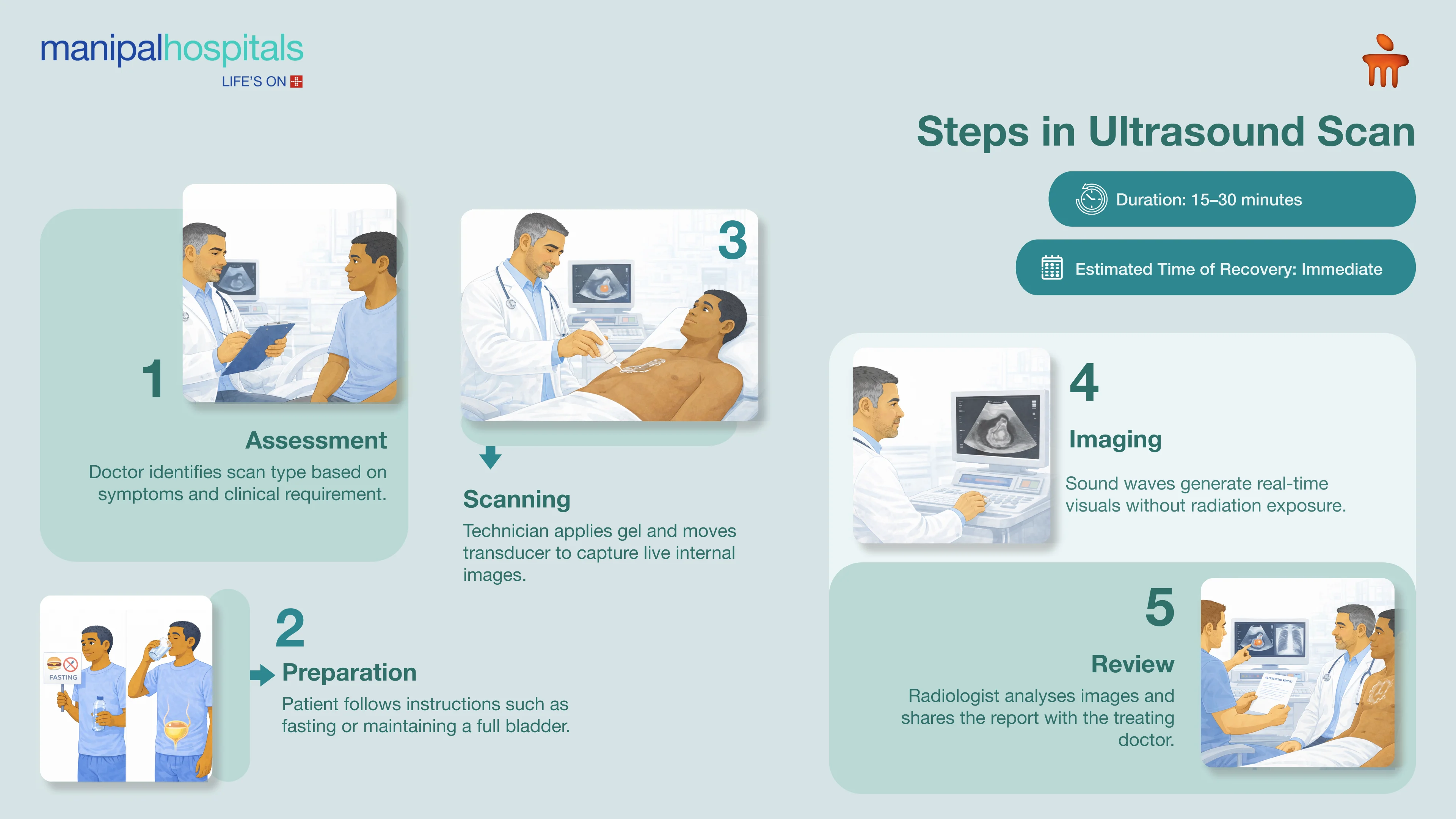
Pregnancy Ultrasound at Manipal Hospitals uses high-frequency sound waves that create real-time images of the baby and uterus without radiation exposure. The process begins with a review of medical history and gestation, helping radiologists choose a transabdominal or transvaginal approach.
For abdominal scans, gel is applied to improve sound transmission before placing the transducer on the abdomen. Early scans may require a transvaginal method, where a slim probe is inserted gently to visualise early structures. Both approaches display images instantly on monitors, allowing clear observation of foetal heartbeat, placenta, amniotic fluid, and anatomical development.
Dating scans (8–11 weeks) assess gestational age using crown-rump length. Nuchal translucency scans (11–14 weeks) help screen chromosomal risks. Anomaly scans (18–24 weeks) examine foetal organs. Growth scans (28+ weeks) use Doppler to assess placental blood flow. Each ultrasound pregnancy test in India provides measurements, movement patterns, and structural details for obstetric review.
A typical scan lasts 15–45 minutes in comfortable rooms. Printed images and digital reports are provided quickly. Clinicians review findings, address concerns, and schedule follow-up scans to support clear communication and continuity of care.
Didn’t find what you were looking for?
Request a CallbackBenefits
Pregnancy Ultrasound at Manipal Hospitals offers several important advantages:
-
Confirms foetal viability, multiple pregnancies, and accurate due dates early.
-
Helps screen chromosomal risks using nuchal translucency assessment.
-
Detects cardiac, skeletal, and organ anomalies for timely intervention.
-
Monitors foetal growth, amniotic fluid, and placental function safely.
-
Supports high-risk pregnancy management without radiation exposure.
What to Expect
Consultation and Diagnosis
Obstetricians review medical history, current symptoms, and previous records to determine the required scans, such as dating, NT, anomaly, or growth assessments.
Pre-operative Preparation
Patients may be advised to maintain a full bladder for abdominal scans to improve clarity, while transvaginal studies require an empty bladder for comfort during an ultrasound pregnancy test in India.
Scan Day
Patients lie comfortably on the exam table. Gel is applied, and the sonographer moves the transducer gently for 20–40 minutes to capture clear foetal and uterine images. For early gestations, a transvaginal probe may be used for better visibility.
Post-Scan Review
No recovery needs apply. Findings are reviewed immediately, with reassurance and explanation provided.
Hospital Stay
Ultrasound is an outpatient procedure, allowing patients to leave soon after completion.
Follow-Up Appointments
Reports are discussed with the obstetric team, guiding any required changes to pregnancy care.
Long-Term Recovery
Serial scans help track development, placental function, and foetal well-being until delivery.
Why Manipal Hospitals?
Manipal Hospitals provides structured pregnancy imaging supported by dedicated Radiology and Foetal Medicine teams. Its ultrasound units use 2D, 3D, 4D, and Doppler technologies designed to assess foetal anatomy, growth patterns, placental health, and blood flow. Radiologists and sonographers interpret scans using standardised protocols that support consistent evaluation across all trimesters. Coordinated pathways link Radiology with Obstetrics, Neonatology, and Foetal Medicine, ensuring that follow-up planning and additional assessments are organised promptly when required. Facilities include private scan rooms, sterile probe systems, and digital reporting platforms that enable clear documentation and timely communication. This integrated approach helps support safe, reliable pregnancy monitoring and guides clinical decisions at each stage of antenatal care.
Centre of Excellence
The Radiology and Imaging Department at Manipal Hospitals performs comprehensive pregnancy ultrasound studies, including dating, NT, anomaly, Doppler, and growth assessments. Advanced imaging systems and skilled teams support accurate foetal evaluation and rapid report delivery. The department works closely with Obstetrics to ensure timely interpretation and integrated antenatal care plans.
Services Offered
Manipal Hospitals provides a range of pregnancy ultrasound services:
-
Viability and dating assessments
-
Nuchal translucency screening
-
Detailed anomaly evaluations
-
Growth and Doppler monitoring
-
3D/4D enhanced imaging
-
Specialised high-risk pregnancy scans
-
Follow-up foetal wellbeing assessments
-
Placental and amniotic fluid evaluation
These services support accurate foetal assessment and structured antenatal care throughout pregnancy.
Facilities & Services
Manipal Hospitals supports ultrasound services with advanced facilities:
-
High-resolution 2D/3D/4D transducers
-
Private, comfortable scan suites
-
Colour Doppler imaging for blood-flow assessment
-
Digital archiving with quick report access
-
Adjacent obstetric support for coordinated care
-
Strict hygiene and sterile probe protocols
-
Temperature-controlled imaging environments
These facilities ensure accurate imaging and patient comfort.
Frequently Asked Questions
An early pregnancy ultrasound confirms the foetal heartbeat, gestational sac location, and viability around 6–8 weeks. It can also identify multiple pregnancies and help provide accurate early dating for planning routine antenatal care.
Ultrasounds use sound waves rather than radiation and are considered safe when medically required and appropriately performed. Scans are recommended only when clinically indicated to assess foetal development, maternal health, and progression across trimesters.
The anomaly scan is typically carried out between 18 and 20 weeks. It evaluates the baby's organs, spine, limbs, heart structures, abdomen, and face to identify developmental concerns and guide further obstetric decisions if needed.
A full bladder improves abdominal imaging by lifting the uterus and creating a clearer acoustic window. Transvaginal ultrasounds, however, are performed with an empty bladder to maximise comfort and achieve early gestational clarity.
If low amniotic fluid is noted, doctors review hydration, medicines, and foetal Doppler flow. Follow-up imaging or a pregnancy ultrasound in India may be advised to track fluid levels, assess placental function, and guide appropriate pregnancy management.
Consult our Experts



You’re on Our Indian Website
Visit the Global site for International patient services
'Click' to chat with us