
DTPA/EC Renogram
Manipal Hospitals provides advanced nuclear medicine imaging to evaluate kidney function, drainage, and perfusion. As a centre for DTPA Renal Scan in India, the hospital offers accurate, safe, and well-monitored assessments performed by trained nuclear medicine specialists. The scan helps measure kidney filtration rate, assess differential function, and support the diagnosis of renal disorders. Using modern gamma cameras and controlled radiopharmaceutical techniques. Manipal Hospitals ensures clear reporting, careful monitoring, and coordinated clinical interpretation to guide further treatment or follow-up planning.
Hospital Locations
-
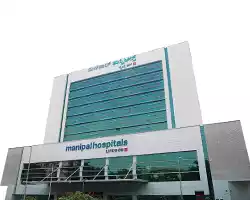
Old Airport Road - Bengaluru
-
.webp)
Mysuru
-
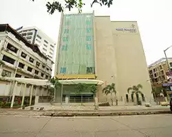
Mangaluru
-
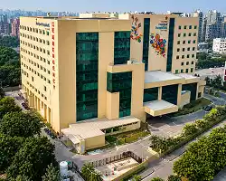
Dwarka - Delhi NCR
-
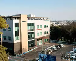
Patiala
-
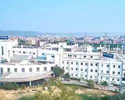
Jaipur
-
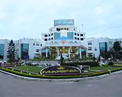
Vijayawada
-
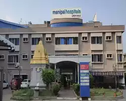
Salem
-
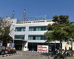
Kharadi - Pune
-
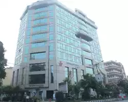
Salt Lake - Kolkata
-
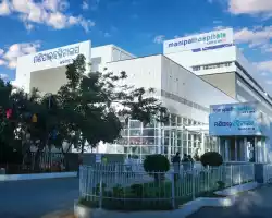
Bhubaneswar
-
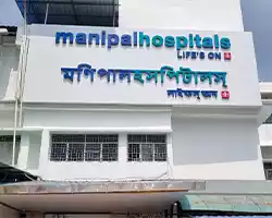
Siliguri
-
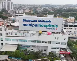
Ranchi
How It Works
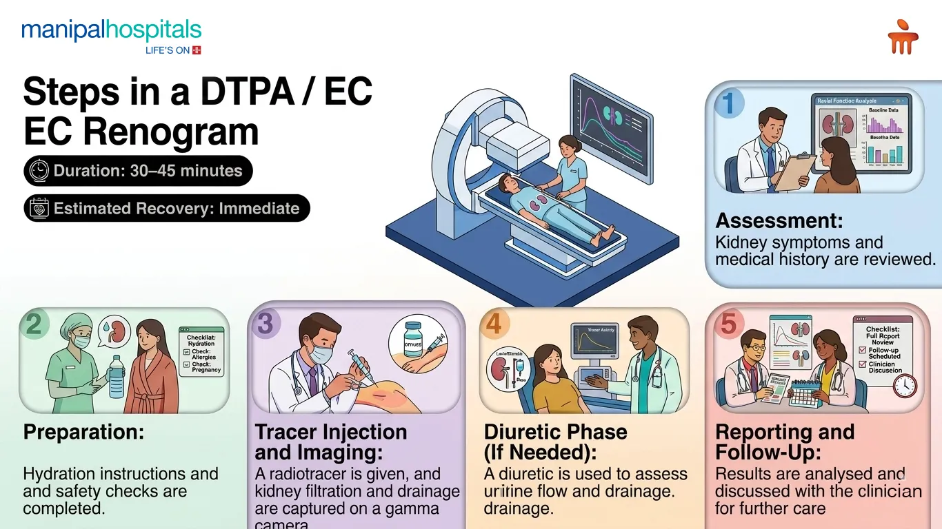
Manipal Hospitals approaches renal imaging through a clear and well-supervised process. It guides patients seeking a DTPA renal scan test in India with structured support at every stage.
Before the procedure, patients undergo a brief clinical review to understand kidney symptoms, previous tests. They may be asked to drink water before arrival to support proper hydration, as this helps radiotracer movement. The care team explains the purpose of the scan, expected sensations, and the approximate timeline.
During the procedure, a small amount of radiotracer is injected into a vein. This tracer travels to the kidneys and allows imaging of their filtration and drainage. Patients lie still while a gamma camera captures continuous images. In some cases, a diuretic may be given to assess urine flow and rule out obstruction. The entire process is monitored closely by trained nuclear medicine specialists to ensure clarity and accuracy.
After the procedure, patients can resume normal activities immediately. Drinking water helps clear the tracer naturally from the body. The results are processed by nuclear medicine physicians who provide a detailed interpretation for the referring doctor. Any abnormalities, obstruction patterns, or differential kidney function results are explained during consultation.
Didn’t find what you were looking for?
Request a CallbackBenefits
Patients receive a detailed, accurate assessment of kidney function through a safe and well-supervised nuclear imaging procedure.
-
Clear evaluation of kidney filtration and drainage
-
Early detection of obstruction or reduced kidney function
-
Non-invasive and painless imaging
-
Useful for monitoring chronic kidney conditions
-
Helpful for pre- and post-surgical assessment
-
Fast results supporting timely treatment decisions
-
Precise differential function measurement for each kidney
What to Expect
Consultation and diagnosis
The process begins with a clinical review of kidney symptoms, medical history, and previous investigations.
Pre-scan preparation
Although no major preparation is needed, patients may be advised to drink water before the scan to enhance tracer movement. They are also asked to inform the team about allergies, pregnancy, or breastfeeding.
Scan day
On the day of the scan, a small dose of radiotracer is injected into a vein. Patients lie on the imaging table while a gamma camera records kidney filtration and drainage in real time.
Post-Scan
After the scan, patients can resume normal activities immediately. Drinking plenty of water helps flush the tracer out of the body.
Follow-up appointments
Doctors discuss the results, highlight any abnormalities, and advise further investigations or treatment if needed.
Why Manipal Hospitals?
Manipal Hospitals is recognised for delivering precise renal imaging supported by advanced nuclear medicine facilities and experienced specialists. The hospital uses high-resolution gamma cameras, controlled radiopharmaceutical protocols, and stringent quality checks. This ensures every DTPA or EC renogram is accurate and dependable. Nuclear medicine physicians, nephrologists, and radiology teams work together to interpret results clearly, helping patients receive timely diagnosis and appropriate treatment recommendations.
Each scan is performed in a well-equipped imaging suite with strict safety measures to minimise radiation exposure and enhance patient comfort. The hospital’s streamlined workflow supports efficient sample handling, rapid image acquisition, and timely reporting. It is vital for evaluating kidney drainage, filtration, and differential function.
Patients benefit from clear pre-scan instructions, guided hydration protocols, and attentive monitoring throughout the procedure. Post-scan discussions help explain findings in simple terms. This approach provides clarity on next steps, such as medication adjustments, further tests, or surgical planning when required.
Centre of Excellence
The Radiology Department at Manipal Hospitals provides advanced imaging solutions for evaluating kidney function and perfusion. Equipped with high-quality gamma cameras and controlled radiopharmaceutical systems, the department delivers accurate DTPA and EC renogram studies. Experienced nuclear medicine physicians ensure safe tracer handling and clear interpretation. With structured workflows and integrated consultation support, the department plays a vital role in diagnosing renal conditions.
Services Offered
Patients receive a complete range of nuclear imaging services designed to assess kidney function with accuracy and comfort.
-
DTPA and EC renogram studies
-
Evaluation of renal filtration and drainage
-
Differential kidney function assessment
-
Diuretic renography for obstruction analysis
-
Imaging for post-surgical or post-treatment monitoring
-
Safe radiotracer administration and guided imaging
-
Detailed interpretation by nuclear medicine specialists
Facilities & Services
Patients benefit from advanced nuclear medicine facilities designed to support safe imaging, accurate reporting, and comfortable scan experiences.
-
High-resolution gamma camera systems
-
Controlled radiopharmaceutical preparation units
-
Dedicated nuclear imaging suites
-
Structured radiation-safety protocols
-
Skilled nuclear medicine physicians and technologists
-
Rapid image processing and reporting
-
Comfortable scan tables for patient ease
Frequently Asked Questions
These scans assess kidney filtration, drainage, and blood flow. They help detect obstruction, measure each kidney’s function, and support diagnosis or treatment planning for various renal conditions.
A DTPA Renal Scan in India at Manipal Hospitals includes a radiotracer injection, gamma camera imaging, filtration and drainage assessment. It also includes a detailed interpretation by nuclear medicine specialists for accurate renal function analysis.
The scan itself is painless. Patients feel only a brief needle prick during radiotracer injection. They lie comfortably while images are captured, and the camera does not cause any pressure, contact, or discomfort.
Results are usually ready the same day or within 24 hours. Nuclear medicine physicians review the images, prepare a detailed report, and share the findings with the treating doctor for further evaluation.
Side effects are uncommon. The radiotracer is given in very small, safe amounts and is cleared naturally through urine. Drinking plenty of water after the scan helps remove it from the body quickly.
Consult our Experts

You’re on Our Indian Website
Visit the Global site for International patient services
'Click' to chat with us