7 Min Read
--How-It-Develops,-Symptoms-and-Treatment-Options.webp "Hepatocellular Carcinoma HCC: Meaning, Symptoms & Treatment Options")
Hepatocellular carcinoma (HCC) is the most common type of primary liver cancer. It begins in hepatocytes, the main functional cells of the liver that handle metabolism, detoxification, and protein synthesis. To answer the common question of what hepatocellular carcinoma (HCC) is – it is a cancer that develops from hepatocytes, the primary functional cells of the liver. It is a cancer that develops within the liver itself and not one that has spread from another organ.
Most cases of HCC are linked to long-standing liver damage. Chronic hepatitis B, hepatitis C, and cirrhosis are the most frequent underlying conditions. Over time, repeated injury to liver cells leads to structural changes that increase cancer risk.
Early disease may not produce clear symptoms. Many patients are diagnosed during routine surveillance for liver disease. This article explains how hepatocellular carcinoma develops, the symptoms to watch for, and the treatment options available.
Synopsis
Hepatocellular Carcinoma Meaning
HCC is the most common type of primary liver cancer, accounting for around 85% to 90% of cases. When people ask what hepatocellular carcinoma means, the term can be broken down simply. “Hepato” refers to the liver, “cellular” to cells, and “carcinoma” to cancer. Together, the term refers to a cancer that originates within the liver cells themselves.
The risk is higher in people with long-standing liver disease. Cirrhosis is the most common underlying condition, although not all cases occur in a cirrhotic liver. In some cases, especially with chronic hepatitis B infection, hepatocellular carcinoma can develop even without cirrhosis.
One of the major challenges with hepatocellular carcinoma (HCC) is that early disease often develops silently without obvious symptoms. In many cases, it gets noticed during regular check-ups for an existing liver issue. That’s why staying consistent with follow-ups is important, especially if someone is at higher risk.
How Hepatocellular Carcinoma Develops
Hepatocellular carcinoma HCC develops over time inside the liver. Repeated liver injury triggers ongoing inflammation and repair within the liver tissue. This repeated cycle leads to inflammation first. Then scar tissue starts forming, which is called fibrosis. If this process continues, the liver becomes cirrhotic. At this stage, the normal structure is lost, and small regenerative nodules appear across the liver.
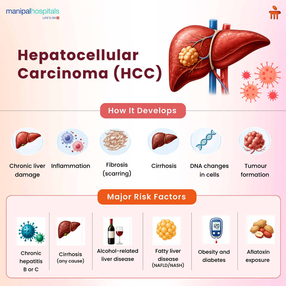
Liver cells are capable of regeneration, but the constant repair is not always accurate. With repeated injury, small errors begin to appear in the DNA of these cells. These abnormal cells begin multiplying uncontrollably despite the body’s normal growth regulation mechanisms.
Over time, these abnormal cells accumulate and form tumours within the liver. These tumours usually have a rich blood supply and can show up as one lump or several, especially in a liver that’s already cirrhotic. As they grow, they begin to affect nearby tissue and gradually reduce how well the liver works.
Risk Factors for Hepatocellular Carcinoma
Most people who develop this cancer already have underlying liver damage, with cirrhosis being the most important risk factor.
Common risk factors include:
-
Chronic hepatitis B or C infection
-
Cirrhosis from any cause
-
Long-term alcohol use
-
Fatty liver disease (including NASH)
-
Obesity and diabetes
-
Exposure to aflatoxins from contaminated food
-
Higher risk in men and older individuals
Hepatocellular Carcinoma Symptoms
Early on, the symptoms of hepatocellular carcinoma can be vague and easy to confuse with other liver issues. In many patients, the disease is detected incidentally during routine surveillance imaging.
Common symptoms include:
-
Pain or heaviness in the upper right abdomen
-
Unexplained weight loss
-
Reduced appetite or early satiety
-
Ongoing fatigue
-
Nausea or general discomfort
-
Yellowing of the skin and eyes (jaundice)
-
Swelling of the abdomen (ascites)
-
Enlarged liver on examination
-
Easy bruising or bleeding
Any sudden worsening of symptoms in someone with cirrhosis should be evaluated promptly.
How Hepatocellular Carcinoma is Diagnosed
Diagnosing hepatocellular carcinoma (HCC) is not based on a single test. It usually builds step by step, especially in patients who are already being followed for liver disease.
Screening in high-risk patients
-
Ultrasound is done every 6 months to check for any new growths in the liver.
-
The AFP (alpha-fetoprotein) blood test may be added, but it is not always accurate on its own.
Diagnostic imaging
-
A contrast CT scan is commonly used once something suspicious is seen
-
MRI is often preferred when doctors need a clearer and more detailed view
-
Doctors look for characteristic enhancement patterns on imaging studies:
-
The lesion picks up contrast quickly in the arterial phase
-
The lesion then demonstrates washout during the venous phase
Biopsy
-
Not required in every case if imaging findings are typical
-
Done when the diagnosis is not clear from scans
Staging and assessment
-
Number and size of tumours present in the liver
-
Liver function is often assessed using the Child-Pugh score
-
Whether the tumour has involved blood vessels or spread outside the liver
Hepatocellular Carcinoma Treatment Options
Treatment for hepatocellular carcinoma HCC is not the same for every patient. The plan depends on how early the tumour is found and how well the liver is functioning. Liver transplant doctors look at the size and number of tumours, where they are located, and whether the rest of the liver is healthy enough to tolerate treatment. The patient’s overall health also matters before deciding on the approach.
Curative Treatments (Early Stage)
-
Surgical Resection: Removal of the tumour along with part of the liver. It is suitable when the disease is limited, and liver function is good. Follow-up is needed as recurrence is possible.
-
Liver Transplant: Replacement of the entire liver with a donor organ. It is considered when there is significant liver damage. Selection depends on tumour size and number, and patients are monitored while waiting for a donor.
Local Ablation Therapies
-
Radiofrequency Ablation (RFA): Uses heat delivered through a needle to destroy tumour cells. It is mainly used for small, localised tumours when surgery is not suitable.
-
Microwave Ablation: Similar to RFA but uses microwave energy for faster and more even heating. It is also used for small tumours.
Locoregional Therapies
-
Transarterial Chemoembolization (TACE): Delivers chemotherapy directly into the tumour’s blood supply and blocks it, reducing blood flow and helping destroy cancer cells. Used when the tumour cannot be removed with surgery.
-
Transarterial Radioembolization (TARE): Uses tiny radioactive particles delivered into the tumour’s blood supply to provide targeted radiation and control tumour growth.
Systemic Therapies (Advanced Stage)
-
Targeted Therapy: Uses medicines such as sorafenib and lenvatinib to slow tumour growth and control spread. These are taken as tablets and require regular monitoring.
-
Immunotherapy: Helps the immune system recognise and attack cancer cells. This treatment is used in selected advanced cases based on the patient’s condition.
-
Supportive and Palliative Care
- Focuses on symptom relief and improving quality of life
- Helps manage pain, fatigue, nausea, and nutritional concerns
- Provides supportive care in advanced stages of disease
Conclusion
Hepatocellular carcinoma needs early attention because it can progress without clear signs. When picked up in time, treatment can focus on removing the tumour. As the stages progress, the focus changes to controlling the disease and managing symptoms. Regular follow-up with top liver transplant hospitals in Sarjapru road and timely decisions make a difference in outcomes. Patients looking for hepatocellular carcinoma treatment in Sarjapur Road, Bangalore, can consult best liver transplantation surgeons near Sarjapur road for proper evaluation, treatment planning, and ongoing care based on their condition.
FAQ's
People with long-term liver problems like cirrhosis, chronic hepatitis B, or C already face a much higher risk. Additional factors like alcohol use or fatty liver disease further increase the risk.
Doctors generally advise a scan every six months. It is an effective way to pick up on early liver changes, often before you feel any symptoms at all.
Not always. It’s often linked to cirrhosis, but it can still happen without it. This condition is more common in people with hepatitis B.
They can help to some extent. Cutting down on alcohol, managing weight, and taking care of existing liver problems may lower the chances over time.
After treatment, doctors usually continue regular follow-ups with scans and tests. This is because there is still a chance that the cancer can come back later.
Share this article on: