9 Min Read
Heart health plays a vital role in overall well-being. Many heart conditions develop silently and show symptoms only when the disease has progressed. Because of this, doctors rely on imaging tests that can evaluate how the heart functions in real time. The echo test is a widely used and non-invasive way to look at the structure and function of the heart. The 2D echo test is a type of ultrasound that lets doctors see the heart's chambers, valves, blood flow, and how well it pumps blood.
When people have chest pain, shortness of breath, palpitations, swelling in the legs, or unexplained fatigue, doctors often recommend the echo heart test. This blog explores how a 2D echo test is performed, what it tells us, and how it may be beneficial for you.
Synopsis
What Is a 2D Echo Test?
A 2D echo test is a type of ultrasound imaging that doesn't hurt and shows the heart's shape and movement. It makes real-time pictures of the heart on a monitor by sending high-frequency sound waves through the heart.
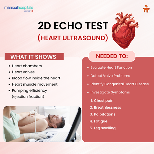
If you think of an echo test as an ultrasound scan just for the heart, it will be easier to understand what is echo test. The test gives doctors two-dimensional pictures that let them look at:
-
Heart chambers
-
Heart valves
-
Blood flow through the heart
-
Thickness and movement of heart muscles
-
Pumping efficiency of the heart
Cardiologists can see how well the heart pumps blood through the body in real time with the echocardiogram. Unlike some other imaging procedures, the echo test does not involve radiation exposure. This makes it safe for repeated monitoring when required.
How Does an Echo Heart Test Work?
Ultrasound technology is used in the echo heart test. A transducer, which is a small device that fits in your hand, sends sound waves into the chest. The waves bounce off the heart's structures and come back as echoes. A computer then turns these echoes into pictures.
The cardiologist can see a number of important things about how the heart works during the 2D echo test:
-
The size and shape of the heart
-
Movement of the heart walls
-
Opening and closing of valves
-
Blood flow patterns inside the heart
-
Possible abnormalities in heart chambers
When people ask what an echo test is, it essentially refers to this ultrasound-based technique that helps doctors evaluate the heart without surgery or invasive procedures.
Because the echo test provides moving images of the heart, doctors can assess both the anatomy and function of the heart in a single examination.
For patients undergoing a 2D echo test in Varthur Road, Bangalore, the procedure typically takes around 20 to 40 minutes and is completely painless.
Why Is a 2D Echo Test Done?
Doctors recommend a 2D echo test for many clinical reasons. It is one of the most effective tools for diagnosing and monitoring heart diseases.
Here are the main situations where an echo heart test may be advised.
1. To Evaluate Heart Function
One of the most common reasons to get an echo test is to see how well the heart pumps blood. The test finds the ejection fraction, which tells you how well the heart pumps blood.
2. To Detect Heart Valve Disorders
Heart valves control how blood moves between the heart's chambers. A 2D echo test can show leaks, valves that are too narrow, or valves that don't open and close the right way.
The echo heart test lets doctors see how the valves are moving in real time, which helps them figure out what's wrong with the heart.
-
Mitral valve prolapse
-
Aortic stenosis
-
Valve regurgitation
3. To Diagnose Congenital Heart Disease
Congenital heart conditions are structural abnormalities present at birth. The echo test is widely used to detect these conditions in both children and adults.
Using the 2D echo test, cardiologists can visualise structural defects such as holes in the heart or abnormal connections between chambers.
4. To Investigate Symptoms
Doctors often recommend an echocardiogram heart test when patients experience symptoms that may indicate heart disease. These include:
-
Breathlessness
-
Chest pain
-
Palpitations
-
Swelling in the legs
5. To Monitor Heart Disease
Patients who already have heart disease may require regular follow-up tests. The 2D echo test is useful for keeping track of how the heart works over time.
Doctors might do the echo test again after surgery, treatment, or changes to medication to make sure the heart is working well.
What Happens During a 2D Echo Test?
If patients know what to expect from the echo heart test, they may feel better about it.
This is what usually happens during the 2D echo test:
-
The patient lies on an examination table.
-
A technician applies a special gel to the chest.
-
A small ultrasound probe is moved across different areas of the chest.
-
Images of the heart appear on a monitor in real time.
-
The technician records several views of the heart.
You don't need to get shots or be sedated for the echo test. Most patients can go back to their normal lives right after the procedure.
Doctors often recommend the test as a routine heart checkup because it doesn't hurt and is safe. This is especially true for people who have risk factors like high blood pressure, diabetes, or a family history of heart disease.
Patients searching for a 2D echo test in Varthur Road bangalore can usually complete the entire procedure within a short hospital visit.
Advantages of a 2D Echo Test
The echo test has become a standard diagnostic tool in cardiology because of its many benefits.
-
Non-invasive: The 2D echo test does not involve surgery or needles, making it comfortable for patients.
-
No Radiation: Unlike CT scans or X-rays, the echo heart test uses sound waves instead of radiation.
-
Real-time Imaging: The echo test provides moving images of the heart, allowing doctors to observe heart function as it happens.
-
Quick and Safe: Most 2D echo test procedures take less than 40 minutes and have no known long-term risks.
-
Accurate Diagnosis: The main advantage is the detailed information it provides about heart structure and function.
For people considering a 2D echo test in Varthur Road bangalore, these benefits make it one of the safest and most reliable heart diagnostic tests available.
Who Should Get an Echo Test?
Doctors may recommend an echo test for individuals with certain symptoms or risk factors.
You may need a 2D echo test if you experience:
-
Irregular heartbeat
-
Persistent chest discomfort
-
Swelling in the legs or ankles
-
High blood pressure
-
History of heart disease
Conclusion
The echo test is one of the most important diagnostic tools used in modern cardiology. It allows doctors to evaluate the structure, function, and overall health of the heart without using invasive procedures. By producing real-time images, the 2D echo test helps detect heart valve disorders, congenital abnormalities, heart muscle damage, and other cardiovascular conditions.
If your doctor has advised a 2D echo test in Varthur Road, Bangalore, timely evaluation can play a crucial role in protecting your heart health. For accurate diagnosis and expert cardiac care, consult the specialists at Manipal Hospital Varthur Road, where advanced diagnostic facilities and experienced cardiologists work together to ensure comprehensive heart evaluation.
FAQ's
An echo test is used to evaluate the structure and function of the heart. It helps detect heart valve disorders, heart muscle problems, congenital defects, and fluid around the heart.
No. The 2D echo test is completely painless and non-invasive. Patients may only feel slight pressure from the ultrasound probe placed on the chest.
A typical echo heart test takes about 20 to 40 minutes, depending on the number of images required.
In most cases, no special preparation is required for a 2D echo test. Patients can eat, drink, and take regular medications unless instructed otherwise.
Yes. The echo test is considered very safe because it uses ultrasound waves rather than radiation.
Doctors recommend an echo heart test to investigate symptoms such as chest pain, breathlessness, irregular heartbeat, or swelling in the legs.
Share this article on:


: Symptoms, Causes, Diagnosis, and Treatment")