7 Min Read
An arteriovenous malformation (AVM) is an abnormal tangle of blood vessels where arteries connect directly to veins, bypassing the capillaries. Capillaries normally slow down blood flow, allowing oxygen and nutrients to be delivered to tissues. In an AVM, this crucial network is missing, leading to high-pressure blood flowing from arteries directly into weaker veins. This can cause the veins to enlarge and potentially rupture, leading to bleeding (haemorrhage) in the brain or other parts of the body.
Synopsis
Where Do AVMs Occur?
AVMs can develop anywhere in the body, but are most found in:
-
Brain
-
Spinal cord
-
Face and scalp
-
Arms and legs
-
Lungs
-
Liver and other internal organs
Brain AVMs are particularly important because they can cause bleeding within the brain.
What Causes AVMs?
The exact cause of an arteriovenous malformation is not fully understood, but most AVMs are believed to be congenital, meaning they are present at birth. They are generally not hereditary, and it is rare for them to be passed down through families. They are thought to arise from an error in foetal development, where the normal formation of blood vessels goes awry. While AVMs are present from birth, symptoms may not appear until later in life, sometimes even decades later, as the malformation grows or complications arise. Acquired AVMs are extremely rare and are usually a result of trauma or infection.
Recognising Arteriovenous Malformation Symptoms
The symptoms of an arteriovenous malformation vary widely depending on its size, location, and whether it has bled. Some individuals with AVMs may never experience symptoms, while others may have severe, life-altering complications. Knowing the symptoms of arteriovenous malformation helps with timely intervention.
Brain AVMs
Patients may experience:
-
Severe headaches
-
Seizures
-
Weakness of an arm or leg
-
Difficulty speaking
-
Visual disturbances
-
Sudden brain haemorrhage (stroke)
Peripheral AVMs (Limbs, Face, Scalp)
Symptoms may include:
-
Swelling
-
Pain
-
Warmth over the affected area
-
Visible pulsations
-
Skin discolouration
-
Bleeding from the lesion
-
Cosmetic deformity
Other AVM Locations
AVMs can also occur in other parts of the body, leading to location-specific symptoms:
-
Pulmonary AVMs (Lungs): Shortness of breath, fatigue, nosebleeds, and reduced oxygen levels.
-
Gastrointestinal AVMs: Chronic bleeding, leading to anemia, fatigue, and blood in stool.
-
Peripheral AVMs (Limbs): Swelling, pain, visible pulsation, and skin discoloration in the affected area.
Diagnosing Arteriovenous Malformations
Early and accurate diagnosis of an arteriovenous malformation helps prevent complications and plan effective arteriovenous malformation treatment. Due to the diverse nature of symptoms, diagnosis often begins with a thorough neurological examination and detailed medical history.
Diagnostic Tools and Procedures
Several advanced imaging techniques are used to accurately identify and characterise AVMs:
-
Magnetic Resonance Imaging (MRI): Via detailed images, MRI helps visualise the AVM's structure and size.
-
Magnetic Resonance Angiography (MRA): A specialised MRI that focuses on blood vessels, revealing the abnormal flow within the AVM.
-
Ultrasound with Doppler: It works by using sound waves to visualise blood vessels (B-mode) while simultaneously measuring the speed, direction, and volume of blood flow (Doppler).
-
Computed Tomography (CT) Scan: Can quickly detect acute bleeding and provide initial structural information.
-
Computed Tomography Angiography (CTA): A CT scan combined with an injected contrast dye to visualise blood vessels and blood flow patterns.
-
Cerebral Angiography (Digital Subtraction Angiography - DSA): In this procedure, a catheter is inserted into an artery (usually in the groin) and guided to the affected organ where contrast dye is injected to provide real-time, highly detailed X-ray images of the blood vessels. This allows experts to map the AVM's feeders, nidus (tangle), and draining veins precisely. This advanced angiography ensures precise arteriovenous malformation treatment planning.
Arteriovenous Malformation Treatment Options
Not every AVM requires immediate intervention. Treatment decisions depend on the location, size, symptoms, risk of bleeding and age and overall health of the patient. The goal is to prevent complications while preserving normal tissue function. Many AVMs are evaluated and managed through Neurological Vascular Intervention procedures, which focus on treating complex vascular abnormalities affecting the brain and nervous system. Some of the options include:
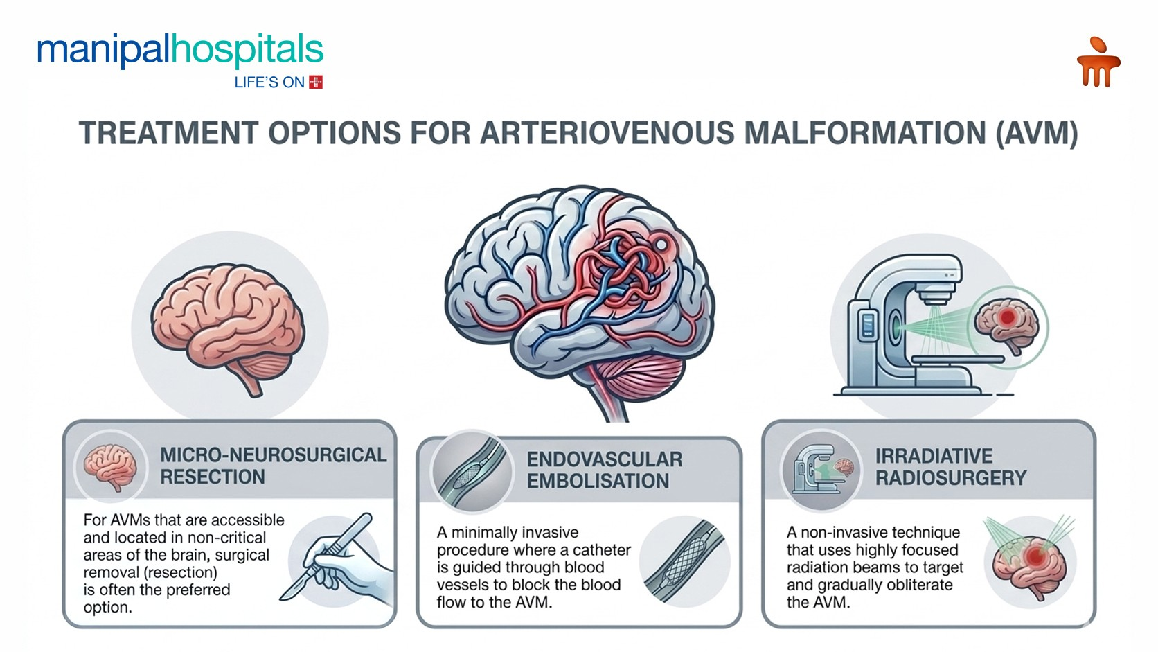
Micro-Neurosurgical Resection
For AVMs that are accessible and located in non-critical areas of the brain, surgical removal (resection) is often the preferred option. This procedure involves opening the skull (craniotomy) to directly visualise and surgically remove the AVM. The goal is complete removal to eliminate the risk of future bleeding.
Endovascular Embolisation
Endovascular embolisation is a minimally invasive procedure performed by an Interventional Radiologist where a catheter is guided through blood vessels to the AVM. A liquid embolic agent (like a medical glue) is then injected into the AVM to block blood flow. This procedure can be used as a standalone arteriovenous malformation treatment, particularly for smaller AVMs, or more commonly, as a pre-surgical step to reduce the AVM's size and blood flow.
Irradiative Radiosurgery
Stereotactic radiosurgery (SRS) is a non-invasive technique that uses highly focused radiation beams to target and gradually obliterate the AVM. It is often used for smaller AVMs (typically less than 3 cm), deeply located, or in critical areas where traditional surgery is too risky.
Combined Approaches and Observation
Sometimes, a combination of these treatments may be used to achieve the best outcome. For example, embolisation might be followed by surgical resection or radiosurgery. In some cases, particularly for very small, asymptomatic AVMs with a low risk of rupture, a "watch-and-wait" approach with regular monitoring might be recommended.
The use of Interventional Radiology has revolutionised AVM management. Many lesions that once required major surgery can now be treated through tiny catheters under image guidance. This approach offers patients, lesser pain, shorter hospital stay, faster return to daily activities, and improved outcomes.
When Should You Seek Medical Attention?
Consult a specialist if you experience:
-
Unexplained swelling or pulsatile masses
-
Recurrent bleeding from a vascular lesion
-
Persistent headaches with neurological symptoms
-
Seizures without an obvious cause
-
Sudden weakness, speech difficulty, or vision loss
Conclusion
Arteriovenous Malformations are uncommon but potentially serious vascular abnormalities. Advances in imaging and minimally invasive endovascular therapies have dramatically improved outcomes. With timely diagnosis and appropriate treatment, most patients can lead normal and productive lives.
If you or a loved one has been diagnosed with an AVM, consultation with a multidisciplinary team, including Interventional Radiology, Neurosurgery, and Vascular Specialists, at Manipal Hospital, Goa, can help determine the most effective treatment strategy.
FAQ's
The most dangerous complication of an AVM is haemorrhage, which can lead to stroke, severe neurological damage, or even be life-threatening.
Most AVMs are congenital (present at birth) but are generally not hereditary, meaning they are rarely passed down through families.
Yes, AVMs can slowly grow larger over time. This growth can increase the risk of rupture and the severity of symptoms.
Recovery time varies greatly depending on the type of treatment, the AVM's location, and whether there were complications. It can range from a few weeks for less invasive procedures to several months or even longer with rehabilitation after a haemorrhage or complex surgery.
After complete surgical resection or successful radiosurgical obliteration, the recurrence of an AVM is rare. However, follow-up imaging is essential to confirm complete eradication and monitor for any new developments.
Since AVMs are typically congenital, lifestyle changes cannot prevent their formation. However, maintaining a healthy lifestyle, especially managing blood pressure, can help reduce the risk of complications if an AVM is present.
Share this article on: