8 Min Read
In the complex journey of cancer diagnosis and treatment, understanding the extent and nature of the disease is paramount. Positron Emission Tomography (PET) scans, often combined with Computed Tomography (CT) scans, offer a powerful window into the body's metabolic activity, providing critical insights that traditional imaging alone cannot. This comprehensive guide aims to demystify the PET CT scan, explaining what is a PET scan, how it works, and when it becomes an indispensable tool in cancer management.
Synopsis
PET CT Scan: How It Works
A PET CT scan is a sophisticated imaging technique that combines two powerful diagnostic methods into one seamless procedure: a PET scan and a CT scan. While a CT scan provides detailed anatomical images, showing the structure and location of organs and tissues, a PET scan reveals metabolic activity at a cellular level. This combination allows physicians to pinpoint areas of abnormal activity and precisely locate them within the body.
The core principle behind a PET scan involves the use of a radioactive tracer, typically a glucose analog like fluorodeoxyglucose (FDG). Cancer cells are known for their rapid growth and higher metabolic rate, meaning they consume glucose much more quickly than healthy cells. When the FDG tracer is injected into your bloodstream, it travels throughout the body and accumulates in areas with high metabolic activity – often cancerous tissues.
The PET scanner then detects the energy emitted by these tracer molecules. A computer processes this information to create detailed, three-dimensional images that highlight these metabolically active areas as bright spots. When fused with the structural images from the CT scan, the PET CT scan provides an incredibly precise map, distinguishing between cancerous and non-cancerous tissues with remarkable accuracy. This integrated approach has been shown to improve diagnostic accuracy by up to 20-30% compared to either scan performed alone, leading to better patient outcomes.
When is a PET scan needed in cancer care?
The utility of a PET scan extends across various stages of cancer care, from initial diagnosis to long-term monitoring. It's not a screening tool for all cancers but is invaluable in specific scenarios where its unique insights are crucial. Here's when a PET scan procedure is typically recommended:
-
Diagnosing Cancer: While biopsies remain the gold standard for definitive diagnosis, a PET scan can help identify suspicious areas that may be cancerous, guiding subsequent biopsy procedures more accurately. It's particularly useful in distinguishing malignant from benign lesions, especially when other imaging methods are inconclusive.
-
Staging Cancer: One of the most critical applications of a PET scan is to determine the extent of cancer spread (staging). It can detect cancer cells that have metastasized to distant organs or lymph nodes, which conventional imaging might miss. Accurate staging is vital for developing an effective treatment plan, with studies indicating PET scans enhance staging accuracy by up to 30% for several types of cancer.
-
Assessing Treatment Effectiveness: During or after chemotherapy, radiation therapy, or other treatments, a PET scan can assess how well the cancer is responding. A decrease in metabolic activity indicates that the treatment is working, while increased activity might suggest resistance or progression. This allows physicians to adjust treatment plans promptly, ensuring optimal care.
-
Detecting Cancer Recurrence: For patients in remission, a PET scan can be highly effective in detecting early signs of cancer recurrence, often before structural changes become apparent on CT or MRI. Early detection of recurrence, which sometimes improves detection rates by 15-20% in asymptomatic patients, can significantly impact prognosis and allow for timely intervention.
-
Radiation Therapy Planning: By precisely delineating the boundaries of tumors and involved lymph nodes, PET scans assist radiation oncologists in planning highly targeted radiation therapy, minimizing damage to surrounding healthy tissues.
-
Identifying the Primary Tumor: In cases of cancer of unknown primary origin (CUP), where metastases are found but the original tumor site is not evident, a PET scan can often locate the primary cancer, guiding further diagnostic and therapeutic efforts.
Benefits of a PET Scan for Cancer Patients
The integration of PET scans into cancer management has revolutionized patient care, offering a multitude of benefits:
|
Comprehensive Whole-Body Imaging |
Unlike many other scans that focus on specific body parts, a PET CT scan can image the entire body in one session, providing a global view of disease activity. This is crucial for detecting distant metastases. |
|
Early Detection |
PET scans can detect cancerous activity at a cellular level, often before structural changes are visible on other imaging techniques. This early detection can lead to earlier intervention and potentially better outcomes. For instance, PET scans can identify lesions as small as 4-5 mm, improving early diagnosis significantly. |
|
Improved Accuracy in Staging |
By identifying all active disease sites, PET scans provide a more accurate staging of cancer, which is fundamental for tailored and effective treatment planning. Researchers observe this increased accuracy in up to 90% of cases for certain cancers like lymphoma. |
|
Personalized Treatment Plans |
The detailed information from a PET scan allows oncologists to create highly personalized treatment strategies, optimizing therapies based on the specific metabolic characteristics and spread of an individual's cancer. |
|
Reduced Need for Invasive Procedures |
By clearly identifying suspicious areas, PET scans can sometimes reduce the need for unnecessary biopsies or other invasive diagnostic procedures, improving patient comfort and reducing risks. |
|
Monitoring Treatment Response |
PET scans are highly effective in monitoring how well cancer is responding to treatment. Within weeks of starting treatment, changes in metabolic activity can indicate efficacy, allowing for prompt adjustments to the treatment regimen. This can lead to better remission rates. |
|
High Patient Safety Profile |
The radioactive tracer used has a very short half-life and quickly exits the body, minimizing radiation exposure. Adverse reactions are extremely rare, occurring in less than 1% of patients. |
Addressing PET Scan Side Effects and Safety
Patient safety is a top priority at Manipal Hospitals. Understanding potential PET scan side effects is crucial, though they are generally minimal and rare.
-
Radiation Exposure: The primary concern for most patients is radiation exposure. It's important to know that the amount of radiation from a single PET CT scan is comparable to other common diagnostic imaging procedures like a standard CT scan. The radioactive tracer used (FDG) has a very short half-life, meaning it decays rapidly and is quickly eliminated from the body, primarily through urine. Our advanced equipment and protocols ensure the lowest possible radiation dose while maintaining diagnostic quality.
-
Allergic Reactions: Allergic reactions to the FDG tracer are extremely rare. Patients are always monitored closely during and after the injection.
-
Discomfort from Injection: Some patients may experience a brief, mild discomfort or a cold sensation at the injection site.
-
For Pregnant or Breastfeeding Individuals: PET scans are generally not recommended for pregnant women due to potential risks to the fetus. Breastfeeding mothers are advised to pump and store milk prior to the scan and avoid breastfeeding for a certain period (usually 12-24 hours) after the scan to prevent exposure to the infant.
Before your PET scan procedure, our medical team will conduct a thorough review of your medical history to ensure your safety and provide specific instructions tailored to your situation.
Conclusion
The PET CT scan stands as a cornerstone in modern oncology, offering an invaluable advantage in the fight against cancer. From its ability to detect cellular activity before structural changes are visible to its precision in staging, treatment monitoring, and recurrence detection, the PET-CT scan provides critical information that empowers both patients and their medical teams. At Manipal Hospitals, we are committed to harnessing this advanced technology, combined with the expertise of our multidisciplinary specialists, to deliver comprehensive, personalized, and compassionate cancer care. Understanding what a PET scan is and its profound impact can significantly influence treatment outcomes and enhance the quality of life for those navigating a cancer diagnosis. We believe in empowering our patients with knowledge and state-of-the-art diagnostics to achieve the best PET scans often enhance disease management, leading to improved health outcomes in over 85% of cases.
If you or a loved one are navigating a cancer diagnosis or treatment journey, understanding advanced diagnostic tools like the PET scan is vital. Our cancer care specialists at Manipal Hospitals Bangalore are dedicated to providing clarity, support, and the highest quality of care.
Book an appointment with us today for a PET scan in Bangalore.
FAQ's
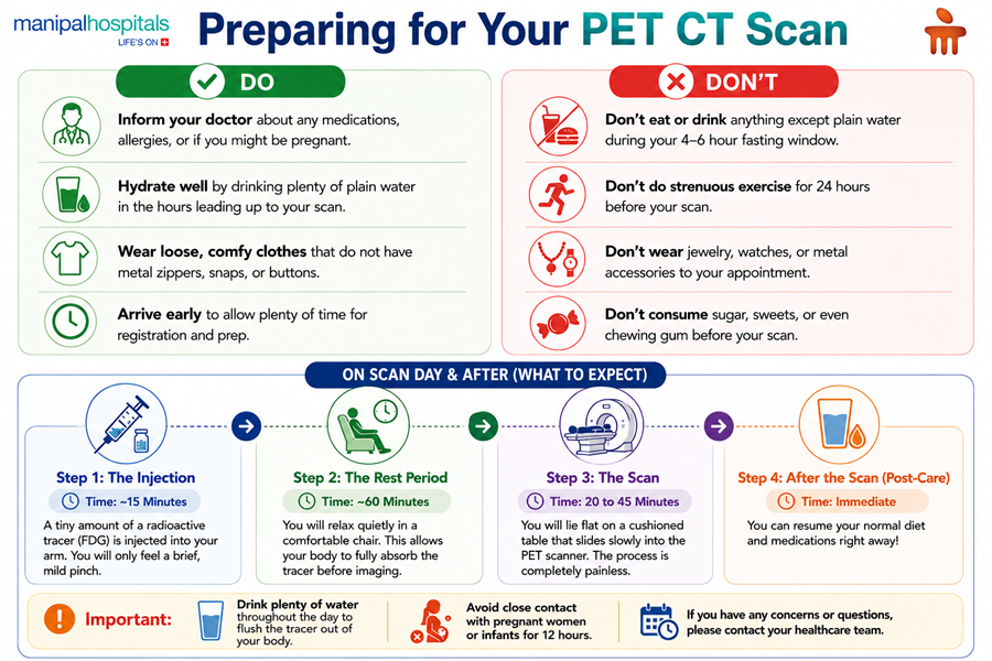
The entire PET scan procedure, from the injection of the tracer to the completion of imaging, typically takes about 2 to 3 hours. The scan itself usually lasts between 20 and 45 minutes.
The procedure is generally painless. You might feel a brief pinprick during the tracer injection. You will lie still on a comfortable table during the scan.
You will typically need to fast for 4-6 hours before your scan, consuming only plain water. Your doctor will provide specific instructions.
The radiation dose from a PET CT scan is carefully controlled and considered safe, comparable to other common medical imaging tests. The tracer rapidly decays and is eliminated from your body.
Our nuclear medicine specialists will interpret your scan, and a report will typically be available to your referring oncologists within 24-48 hours. Your doctor will then discuss the results with you.
Share this article on: