6 Min Read
For many women, the word mammogram brings a level of concern. But it is a highly effective way to detect breast cancer early and identify changes in the tissue long before symptoms appear. Despite mammograms being proven to save lives, questions still arise, especially about cancer risk from repeated screening and radiation from mammograms.
In view of Breast Cancer Awareness Month, this blog aims to separate facts from misconceptions and explain when and why a doctor may recommend a mammogram. We discuss what the test involves, the level of exposure it carries, and how medical guidelines balance safety with necessity. Understanding the real information behind mammogram cancer risk can help women make informed decisions with confidence.
Synopsis
What is a Mammogram?
A mammogram is a low-dose X-ray imaging test that allows doctors to examine breast tissue for early signs of cancer. It is quite an effective way for early detection of cancer, as it can make it possible to detect changes before any symptoms appear. Detecting cancer early gives better chances for treatment and recovery. Hence, understanding mammogram cancer risk helps patients approach screening with clarity rather than fear.
Types of Mammograms
Mammograms are broadly divided into two types:
-
Screening Mammogram: Done as a routine check-up for women without symptoms. It helps detect small lumps or tissue changes that may not be felt during a physical exam.
-
Diagnostic Mammogram: Recommended when there is a lump, pain, or abnormal result from a screening test. It involves more detailed images for accurate evaluation.
Both involve very low radiation from mammograms and are performed under safe, regulated protocols.
What Are 3D Mammograms And When Are They Recommended?
Unlike a standard mammogram which produces only 2D images, a 3D mammogram produces both 2D and 3D images. When used together for breast cancer screening, it can significantly improve detection in individuals with dense breast tissue, as it allows doctors to visualise the regions beyond the areas of density.
Dense breast tissue generally comprises milk glands, milk ducts and supportive breast tissue. It is different from fatty tissue, which appears dark or transparent on a regular mammogram. On the other hand, both dense tissue and breast cancer appear white, making it hard to tell them apart. This is where a 3D mammogram comes handy. Furthermore, they also reduce the need for follow-up imaging.
How Mammograms Are Performed
During a mammogram, the breast is placed on a flat plate while another plate presses gently from above. This compression spreads the tissue evenly, so clear and detailed X-ray images can be taken from different angles. The procedure is fast and is over in a few minutes.
Some women may experience mild discomfort or pressure during compression, which is normal. The short duration and careful technique make it safe for most patients. The level of radiation from mammograms is kept very low and within medical safety standards. Understanding this process helps reduce anxiety.
Understanding the Risk of Radiation
Many women worry that repeated mammograms might increase their cancer risk because the test uses ionising radiation. In reality, the amount used in mammography is very low and carefully controlled. The radiation is just enough to create a clear image of breast tissue, and far below levels known to cause harm.
How Much Radiation Is Involved
The amount of radiation from mammograms is extremely small. One test puts the body at about the same radiation exposure a person gets from nature for a period of two to three months. Even after many years of constant screening, the extra exposure is still considerably small. All medical imaging complies with very stringent safety regulations, so the doses remain far below the levels that can cause harm.
Balancing Risk and Diagnostic Need
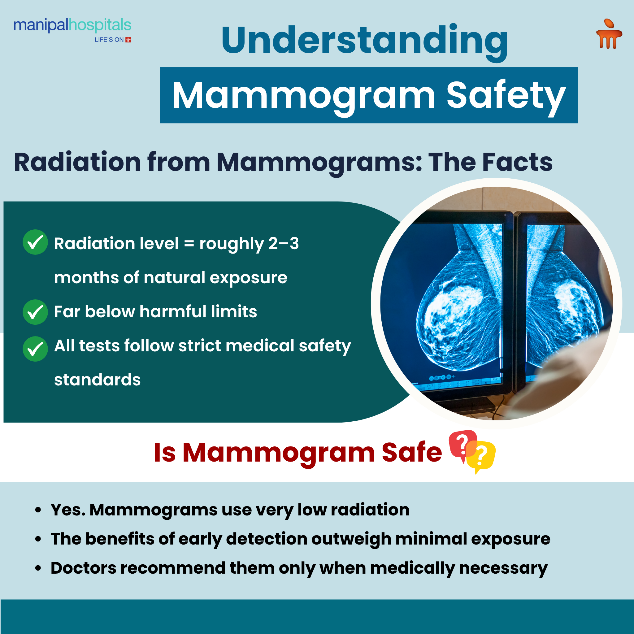
Many women ask, “Is a mammogram safe?” It must be stated that any X-ray procedure carries a slight increase in the risk. But the benefit of early breast cancer detection far outweighs the risk that comes from scans. Missing an early diagnosis can have much greater consequences than the controlled radiation exposure from screening. Doctors recommend mammograms only when there is a clear medical need. The test is only recommended after considering each woman’s age, health history, and personal risk factors. Following medical guidance is the safest approach to managing mammogram cancer risk responsibly.
Who Should Get a Mammogram?
They are prescribed based on a woman’s medical history and overall risk profile. The goal is to balance safety and necessity while reducing mammogram cancer risk through timely detection.
Doctors usually consider the following factors:
-
Age: Women aged 40 years and above are usually advised to undergo regular mammogram screening every one to two years.
-
Family or genetic risk: Those with a family history of breast cancer, known genetic mutations (like BRCA1 or BRCA2), or past chest radiation may need earlier or more frequent tests.
-
Personal medical history: Women with dense breast tissue or previous abnormalities might be advised to undergo additional imaging, such as an ultrasound or MRI.
-
Doctor’s recommendation: The decision on when to get mammogram screening and how often should always come from a qualified medical professional familiar with the patient’s history.
Conclusion
The mammogram cancer risk linked to repeated screenings is very small. It is more so when compared to the life-saving value of early breast cancer detection. Mammograms use low levels of radiation and are performed only when medically justified. Avoiding this test out of fear can delay diagnosis and reduce treatment options for breast cancer that might have been detected earlier.
Doctors recommend mammograms after carefully assessing each woman’s risk factors, health status, and age. The safest approach is to trust your doctor’s judgment and maintain open discussions about when to get mammogram screening and how often it should be repeated.
For women seeking expert evaluation and breast health guidance, Manipal Hospital Old Airport Road, Bangalore, offers advanced diagnostic imaging under the supervision of experienced specialists.
FAQ's
Repeated mammograms expose you to a small amount of radiation. But the exposure is minimal and considered safe. It is understood that the benefit of early detection of cancer far outweighs the mammogram cancer risk.
Mammograms are safe even for women with dense breast tissue. But in such cases, other scans such as ultrasound may be used for further detection.
The frequency depends on age, health history, and risk factors. When to get mammogram screening is best determined by your doctor. Many women aged 40 and above are advised to screen every one to two years.
The compression of the breast during the test does not cause cancer cells to spread. The radiation from mammograms also does not cause the cancer to spread. So there’s nothing to worry.
Women above the age of 40 are recommended to undergo the scan. But women with a family history or higher genetic risk may need to start earlier. Always ask for your doctor’s suggestion for what is best.
Share this article on:


2.png)