7 Min Read
What looks like a small sore on the foot can quietly become the most severe problem a person with diabetes faces. A seemingly minor injury can evolve into a serious infection or even lead to amputation when diabetes affects the feet. That progression is not inevitable, but understanding why wounds get stuck, how to spot danger early, and which treatments actually work can change outcomes. This blog explains the biology behind diabetic foot problems, the signs families should never ignore, and the practical steps clinicians take to reduce amputation risk and preserve mobility.
Synopsis
- Diabetes and Foot Ulcers
- Are You at Risk of Diabetic Foot Ulcers?
- Early Warning Signs Families Should Never Ignore
- How Doctors Assess a Diabetic Foot?
- Why Some Ulcers Become Non-Healing
- Principles of Effective Diabetic Foot Treatment
- Common Treatments and When They Matter
- Prevention: Daily Practices That Save Feet
- When Amputation Becomes Necessary?
- Conclusion
Diabetes and Foot Ulcers
The foot is a common site of chronic wounds and infection in diabetes patients. They are mainly vulnerable to foot problems for 3 major reasons. These are:
-
Loss of protective feeling from peripheral neuropathy: Small cuts or pressure points go unnoticed and continue to stress the feet
-
Poor blood flow from peripheral arterial disease: Tissues lack the oxygen and nutrients needed to repair themselves
-
Impaired immune and healing responses related to prolonged high blood sugar: The body struggles to fight infection and mount normal healing
Over time, these problems create a cycle: an unnoticed blister breaks down into a diabetic foot ulcer, bacteria colonise the wound and form a stubborn biofilm, the wound fails to progress through normal healing stages, and infection may spread into deeper tissues, including bone. That sequence is why clinicians treat foot sores in diabetes far more aggressively than similar minor wounds in people without diabetes.
Are You at Risk of Diabetic Foot Ulcers?
A substantial share of people with diabetes will develop a foot complication during their lifetime, and prior ulceration or amputation is the strongest predictor of recurrence. Additional risk factors include long-standing diabetes, poor blood sugar control, smoking, peripheral neuropathy, visible foot deformity, and reduced pedal pulses. Regular foot checks help clinicians stratify risk and schedule preventive care.
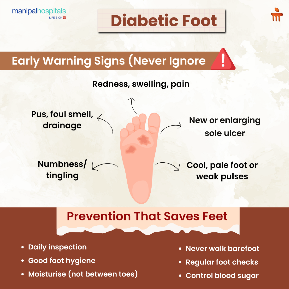
Early Warning Signs Families Should Never Ignore
A timely clinical review can stop progression. Families and carers should pay attention to the following early signals:
|
Warning Sign |
What It May Mean |
|
Redness, swelling, increasing pain |
Local infection or spreading cellulitis |
|
Drainage, foul smell, or pus |
Bacterial colonisation or deep infection |
|
New or enlarging sore, especially on the sole |
Pressure point breakdown (risk for diabetic foot ulcer) |
|
Numbness or tingling |
Peripheral neuropathy and loss of protective sensation |
|
Cool, pale foot or weak pulses |
Peripheral arterial disease and poor perfusion |
If any of these are present, an urgent clinical review is the safest step; early intervention prevents many admissions and reduces amputation risk.
How Doctors Assess a Diabetic Foot?
A structured clinical assessment is the foundation of good diabetic foot care. Typical elements include:
-
A detailed wound inspection (depth, presence of slough, exposed tendon or bone).
-
Neurological testing for protective sensation (monofilament or vibration).
-
Vascular assessment, including palpation of pulses and bedside measures such as the ankle-brachial index or toe pressures.
-
Microbiological sampling when infection is suspected (tissue sampling preferred over superficial swabs).
-
Imaging a deeper infection (osteomyelitis) or bone involvement is suspected (X-ray, MRI).
-
Systemic evaluation of glucose control, nutrition, and other comorbidities that hinder healing.
This broad approach avoids treating only the surface problem and instead aims to correct the factors that keep a wound from healing.
Why Some Ulcers Become Non-Healing
A non-healing foot ulcer results when the balance between tissue damage and repair favours damage. Important drivers include:
-
Repeated pressure and shear at the same spot (particularly the plantar surface)
-
Ongoing infection or bacterial biofilm that resists routine treatment
-
Critical limb ischemia from peripheral arterial disease that prevents tissue oxygenation
-
Poor glycaemic control and malnutrition impair cellular repair
-
Structural deformity (Charcot foot) that alters pressure distribution
Addressing each of these domains is essential to convert a stagnant ulcer into a healing wound.
Principles of Effective Diabetic Foot Treatment
Treating foot complications in diabetes follows a logical sequence: rapid infection control, rigorous wound bed preparation, relieving pressure, restoring blood flow when needed, and optimising systemic health. These steps are standard in international guidance and form the backbone of modern wound care for diabetic feet.
Infection Control: Treat the Wound, Not Just the Swab
Not every colonised ulcer needs systemic antibiotics; only signs of infection or spreading cellulitis do. Tissue cultures guide targeted therapy in deep or persistent infections. When infection reaches bone (osteomyelitis), combined medical and surgical management is frequently required. Delaying treatment increases the chance of limb-threatening spread and, therefore, higher amputation risk.
Wound Debridement: Achieve Healthier Feet
Removing necrotic tissue and biofilm is fundamental. Wound debridement, sharp surgical or regular clinical debridement, is considered a cornerstone to create a healthy wound bed and is recommended repeatedly in guidance documents. Debridement also reduces bioburden and improves the effect of dressings and topical agents.
Offloading: The Most Underused Yet Powerful Tool
Offloading (taking pressure off the ulcer) is frequently the single most effective intervention for plantar ulcers. A properly fitted total contact cast or a pressure-reducing walker removes the repetitive trauma that prevents healing. When offloading is not feasible, therapeutic footwear and custom insoles reduce recurrence risk once the ulcer has healed. Here, patient adherence is the key: the most effective device is the one the person will wear consistently.
Revascularisation: When Circulation Is the Limiting Factor
If vascular studies show severe ischemia, restoring blood flow through angioplasty, stenting, or bypass surgery is often essential to prevent amputation and save the limb. Revascularisation converts a hostile, hypoxic wound bed into one capable of healing, and multidisciplinary vascular input should be early when ischaemia is suspected. Revascularisation enables subsequent wound healing by restoring oxygen and nutrient delivery. Multidisciplinary vascular assessment is essential in ischaemic ulcers.
Each element supports the others: for example, debridement without offloading often fails because the pressure continues to injure the wound.
Optimise Systemic Health
Good glycaemic control, nutrition, smoking cessation, and management of comorbidities are non-negotiable. Coordinate with your primary care team to create a holistic plan; wound care alone is rarely enough.
Common Treatments and When They Matter
|
Treatment |
Indication |
What It Does |
|
Debridement (sharp/mechanical) |
Necrotic tissue or slough |
Removes devitalised tissue and reduces biofilm |
|
Systemic antibiotics |
Clinical infection, spreading cellulitis, osteomyelitis |
Treats bacterial infection that can otherwise progress |
|
Offloading (total contact cast, removable walker) |
Plantar pressure ulcers |
Redistributes pressure to allow granulation and closure |
|
Revascularisation (endovascular/surgical) |
Critical limb ischemia, poor perfusion |
Restores blood flow so tissue can heal |
|
Advanced dressings / NPWT |
Large exuding wounds after debridement |
Manages exudate and promotes granulation |
|
Glycaemic optimisation & nutrition |
Poor metabolic control or malnutrition |
Improves cellular repair and infection resistance |
Prevention: Daily Practices That Save Feet
Most serious outcomes are preventable when people with diabetes and their carers adopt consistent foot care habits. Daily inspection, basic foot hygiene, protective footwear, prompt treatment of minor injuries, and regular professional foot checks are the pillars of prevention. Education must be practical: advise checking between toes, avoiding barefoot walking, cutting nails straight across, and reporting any change immediately.
-
Inspect feet daily for cuts, blisters, redness, or swelling.
-
Keep skin moisturised, but avoid cream between toes.
-
Wear well-fitting shoes and avoid walking barefoot.
-
Have a professional foot exam at least annually, more often if high risk.
-
Keep blood glucose and other chronic conditions well controlled.
These measures lower the chance that a minor injury will become a life-changing problem.
When Amputation Becomes Necessary?
Despite best efforts, some limbs cannot be salvaged. Amputation decisions consider the extent of infection, the chance of functional recovery, comorbidity burden, and patient preferences. The primary aim is to remove infected, nonviable tissue while preserving as much function as possible. Early, limb-sparing interventions combined with rehabilitation often result in better quality of life than delayed major amputation following prolonged sepsis. Multidisciplinary discussion and shared decision-making are essential in these difficult scenarios.
Conclusion
Diabetes affects the feet through a combination of nerve damage, poor circulation, and impaired healing. Early detection, simple daily foot hygiene, pressure relief with offloading, targeted infection control, and timely revascularisation when needed dramatically reduce amputation risk. Most importantly, coordinated care from a multidisciplinary team and clear patient education at Manipal Hospitals convert threats into manageable risks. With attention and the right interventions, most people with diabetes can keep their feet healthy and mobile.
FAQ's
At least once a year for most people; more frequently (every 1–3 months) if they have loss of sensation, prior ulcers, deformity, or circulation problems.
They should stop putting pressure on the area, keep it clean, avoid home remedies that can delay care, and seek medical review promptly. Early action prevents many complications.
Yes. Better glycaemic control improves healing and reduces infection risk; it is a key component of the overall wound care strategy.
Therapeutic footwear and insoles reduce recurrence by redistributing pressure. In people at high risk, pressure-optimised footwear is recommended.
Not always; many infections respond to antibiotics and debridement. Surgery is necessary when there is deep infection, extensive necrosis, or osteomyelitis that does not respond to conservative measures.
Share this article on: