7 Min Read
An open fracture is one of the most urgent injuries an orthopaedic surgeon sees. Unlike a broken bone that stays hidden beneath intact skin, an open fracture breaks through the skin’s protective barrier, leaving the bone exposed to the outside environment. The moment that barrier is lost, the risk shifts from a straightforward bone healing process to one complicated by potential infection, soft tissue damage, and the need for precise surgical reconstruction.
While the thought of a bone piercing the skin is frightening, it is important to know that modern orthopedic care has transformed the outlook for these injuries. With early, systematic open fracture management, most patients go on to heal successfully and return to daily life.
In this article, expert insights from a top orthopaedic hospital in Mangaluru help you learn about what is an open fracture, the common types, the potential open fracture complications, and what to expect during recovery.
Synopsis
Understanding Open Fractures and How They Occur
Inside the body, bones are surrounded by layers of muscle, fat, and skin that protect them from contamination. An open fracture, sometimes called a compound fracture, occurs when a broken bone pierces through these layers or a wound reaches down to the bone. This connection between the break and the outside world allows dirt, debris, and bacteria to reach deep tissues, making infection a primary concern from the very first moment of injury.
Open fractures typically happen through high-energy events. Road traffic accidents, falls from a height, and industrial mishaps are among the most common causes. The shinbone sits just under the skin along the front of the leg and is particularly vulnerable. The surrounding muscle, blood vessels, and nerves can also sustain significant damage. This is why an open fracture is approached as a limb-saving emergency rather than just a fracture that happens to have a wound.
Common Open Fracture Types
Orthopaedic surgeons classify open fractures to guide treatment and predict outcomes. Understanding the open fracture types helps explain why some injuries require a single surgery while others need multiple procedures. Common types according to the Gustilo-Anderson classification include:
|
Open Fracture Type |
Description |
|
Type I |
A small wound, usually less than a centimetre, where the bone has pierced the skin from the inside. The soft tissue damage is minimal, and the wound is relatively clean. |
|
Type II |
A wound larger than a centimetre with moderate soft tissue injury. There is more bruising and muscle damage, but the bone can usually be covered without complex reconstruction. |
|
Type III A |
A high-energy injury with extensive soft tissue damage, yet there remains enough healthy tissue to close the wound directly. |
|
Type III B |
Extensive soft tissue loss where the bone is stripped of its covering. The wound cannot be closed without a flap of tissue moved from another area to cover the exposed bone. |
|
Type III C |
The most severe type, involving major blood vessel injury that compromises circulation to the limb. This requires vascular repair alongside fracture and wound management. |
Common Open Fracture Symptoms
An open fracture is often immediately recognisable because the broken bone may be visible through a wound in the skin. Common signs include severe pain, significant bleeding, swelling, bruising, and a noticeable deformity of the affected limb.
Patients may find it difficult or impossible to move or bear weight on the injured area. In more serious cases, numbness, tingling, pale skin, or a cold hand or foot below the injury can occur, indicating possible nerve or blood vessel damage that requires urgent medical attention.
Immediate Management: What Happens in the Emergency Room
Open fracture treatment starts the moment the patient is picked up by emergency services and continues intensively in the hospital. The early steps are critical in reducing the risk of open fracture complications.
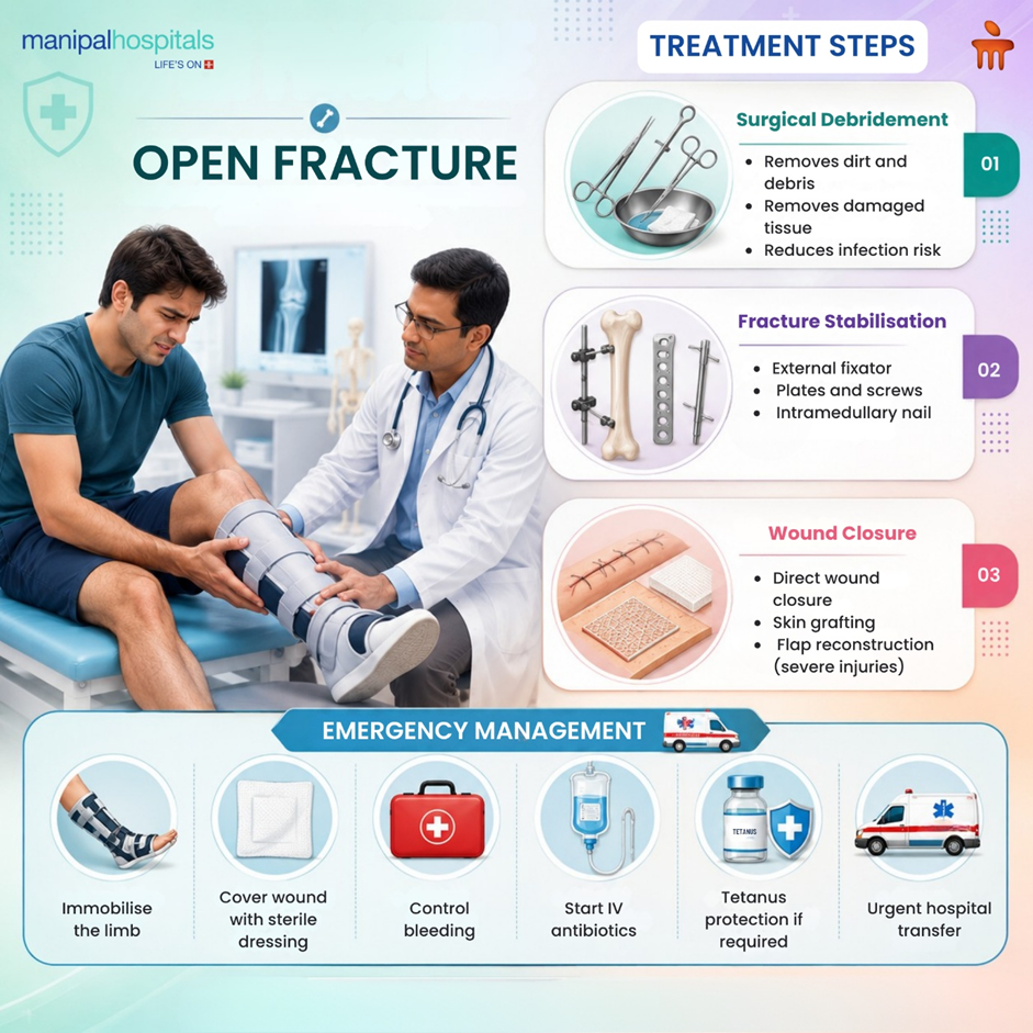
Immediate measures include:
-
Splinting the limb in the position found to prevent further soft tissue damage
-
Covering the wound with a sterile saline-soaked dressing
-
Starting broad-spectrum intravenous antibiotics immediately
-
Administering a tetanus booster if immunisation status is unclear
-
Assessing the blood supply and nerve function of the limb
-
Arranging urgent X-rays and sometimes a CT scan for surgical planning
In the emergency room, the orthopaedic team evaluates the wound, the fracture pattern, and the patient’s overall condition. The operating theatre is prepared as soon as possible because the time from injury to thorough surgical cleaning is one of the strongest predictors of avoiding deep infection.
Surgical Treatment: Debridement, Stabilisation, and Closure: Surgery for an open fracture has several distinct objectives, and all must be achieved systematically to give the limb its best chance of healing well.
-
Surgical debridement and irrigation: In the operating room, the wound is extended and explored. All dirt, foreign material, and dead or dying tissue are meticulously removed. Copious amounts of sterile fluid are used to wash the wound. This debridement is the bedrock of open fracture management; if dead tissue is left behind, bacteria will thrive regardless of antibiotics.
-
Bone stabilisation: Once the wound is clean, the fracture must be stabilised. Depending on the injury, the surgeon may use an external fixator, where pins are placed into the bone above and below the fracture and connected by bars outside the skin. This is often the choice for heavily contaminated or unstable soft tissue injuries. In less severe open fractures, internal fixation with plates and screws or an intramedullary nail can be performed at the same sitting.
-
Wound closure and soft tissue coverage: If the wound can be closed without tension, this procedure is often done at the initial surgery or after a second-look debridement 48 hours later. For wounds with significant tissue loss, a temporary dressing is placed, and a plastic surgeon steps in to provide coverage with a skin graft or a flap of muscle and skin moved from a nearby area. Closing the wound safely is essential; a closed wound protects the bone from ongoing contamination.
-
Antibiotic therapy: Intravenous antibiotics started in the emergency room are continued after surgery. The duration and choice of antibiotics depend on the open fracture types and the degree of contamination. This is a carefully tailored part of open fracture treatment to minimise infection risk.
Potential Open Fracture Complications
Even with excellent surgical care, open fractures carry a risk of complications that patients should know about. Early recognition improves outcomes. The main open fracture complications include:
-
Infection: Superficial wound infections can be treated with antibiotics and dressings. Deep bone infection (osteomyelitis) is more serious and may require further surgery and prolonged antibiotics.
-
Delayed union or non-union: The bone may take longer than expected to heal, or it may fail to heal altogether. Such cases can require bone grafting or revision fixation.
-
Malunion: The bone heals in a poor position, potentially affecting limb function and alignment.
-
Nerve or blood vessel damage: Some residual numbness or weakness may persist if nerves were injured at the time of trauma.
-
Compartment syndrome: Swelling within the muscle compartments of the limb can build up dangerous pressure, requiring an emergency release procedure called a fasciotomy.
-
Stiffness and loss of function: Prolonged immobilisation can lead to joint stiffness and muscle wasting, which require dedicated rehabilitation.
What to Expect During Open Fracture Recovery
Recovery from an open fracture is a gradual, multi-phase process that extends well beyond the initial hospital stay. During the early phase, the focus is on wound healing and controlling pain. The limb may be elevated, and movement is restricted based on the stability of the fixation. Physiotherapy begins gently, often with exercises for the joints that are not immobilised, to prevent stiffness. As healing progresses, weight-bearing is introduced according to the surgeon’s instructions. Loading the bone too early can jeopardise fixation and healing.
Successful open fracture treatment recovery depends on:
-
Following weight-bearing restrictions precisely
-
Keeping the wound and pin sites clean and dry
-
Attending all physiotherapy appointments and doing home exercises consistently
-
Eating a diet adequate in protein, calcium, and vitamin D to support bone healing
-
Avoiding tobacco, which significantly impairs fracture healing
-
Reporting any increase in pain, drainage, redness, or fever promptly
Full recovery, including return to physically demanding work or sport, can take six months to a year or longer depending on the severity of the initial injury and the presence or absence of open fracture complications.
Conclusion
An open fracture is a serious orthopedic injury, but it is one for which clear, evidence-based treatment pathways exist. Understanding what is an open fracture and the rationale behind urgent surgical debridement, stabilisation, and soft tissue coverage makes the journey less daunting.
If you or a loved one needs expert care, comprehensive fracture treatment in Mangalore at KMC Manipal Hospital, consult our experienced orthopaedic surgeons in KMC Manipal Hospital. They ensure both surgical precision and personalised recovery support.
FAQ's
Yes. The exposure of bone and deep tissues to the environment means that infection risk is high from the moment of injury. Surgical cleaning and stabilisation should occur as soon as possible, ideally within 24 hours, to minimise this risk.
An external fixator allows rapid stabilisation with minimal additional soft tissue disruption. It is often the preferred choice when there is heavy contamination, extensive soft tissue swelling, or when the patient is not stable enough for a longer internal fixation procedure.
Many open fractures heal without bone grafting. However, if there is significant bone loss at the time of injury or if healing stalls, a bone graft may be performed later to stimulate the healing process.
Warning signs include increasing pain, redness or warmth around the wound, thick or foul-smelling drainage, and fever with chills. Any of these symptoms should be reported to your surgical team immediately.
Many patients regain functional movement that allows them to perform daily activities and return to work. The extent of recovery depends on the severity of the original soft tissue injury, the joints involved, and commitment to physiotherapy. Some residual stiffness or mild weakness is possible, especially after high-grade injuries.
Share this article on:


 Surgery")