7 Min Read
The human heart is a remarkable and complex organ that works continuously to pump blood throughout the body. To keep it functioning properly, early detection of heart problems is extremely important. When doctors evaluate heart health, one of the most commonly recommended tests is echocardiography.
Many patients, however, feel confused when they hear terms like 2D echocardiography and 3D echocardiography. What do they mean? Are they different tests? And which one is better?
At Manipal Hospital Kharadi, Pune, cardiologists use advanced imaging technologies to diagnose heart conditions accurately and guide treatment decisions. Understanding the difference between 2D and 3D echocardiography can help patients feel more confident and informed about their cardiac care.
Synopsis
- Understanding Echocardiography: A Window Into Your Heart
- 2D Echocardiography: The Standard and Reliable Heart Scan
- 3D Echocardiography: Advanced Cardiac Imaging
- 2D vs 3D Echocardiography: Which Test is Right for You?
- Care Essentials: Do's and Don'ts
- Expert Cardiac Care at Manipal Hospital Kharadi
- Conclusion
Understanding Echocardiography: A Window Into Your Heart
What is Echocardiography?
Echocardiography, commonly called an echo test, is a non-invasive imaging procedure that uses ultrasound waves to create real-time pictures of the heart. These sound waves bounce off heart structures and produce detailed moving images that help doctors observe how the heart is functioning.
This test allows cardiologists to examine:
-
Heart chambers
-
Heart valves
-
Blood flow patterns
-
Heart muscle strength
-
Surrounding blood vessels
The best part is that echocardiography does not use radiation, making it a safe diagnostic tool for people of all ages.
Why is Echocardiography Important?
An echocardiogram plays a crucial role in diagnosing and monitoring many heart conditions. Doctors often recommend it to evaluate:
-
Heart valve diseases
-
Heart failure
-
Congenital heart defects
-
Heart muscle weakness
-
Blood flow abnormalities
-
Structural heart problems
Cardiologists also compare the results with echocardiography normal values to determine whether the heart is functioning within a healthy range. Early detection through echocardiography often allows doctors to start treatment sooner and prevent complications.
2D Echocardiography: The Standard and Reliable Heart Scan
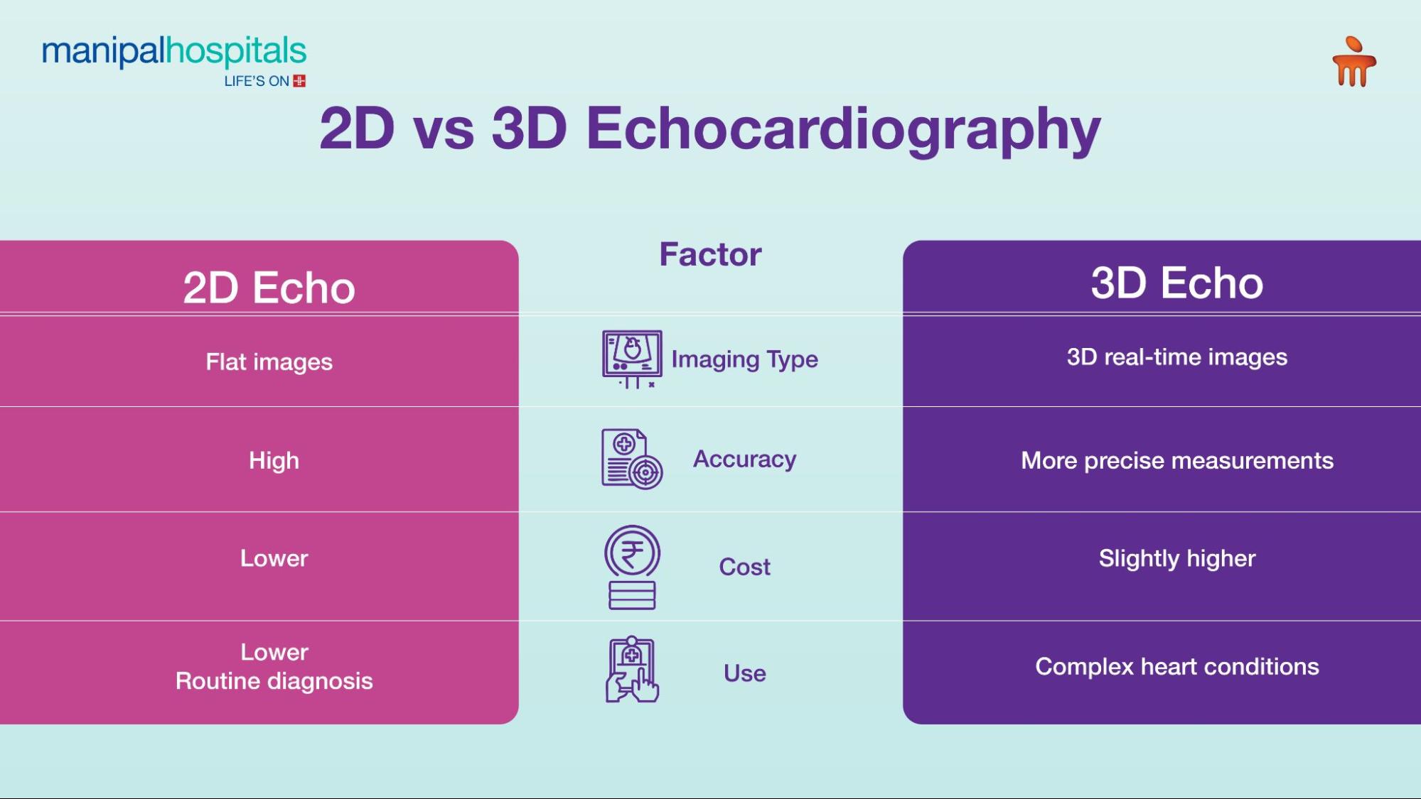
For many years, 2D echocardiography has been the primary imaging technique used in cardiology. It produces two-dimensional cross-sectional images of the heart.
You can think of it like viewing slices of the heart from different angles, similar to flipping through pages of a book.
Benefits of 2D Echocardiography
1. Widely Available
2D echo machines are available in most hospitals and cardiac clinics, making it a commonly used diagnostic tool.
2. Cost-Effective
Compared with advanced imaging techniques, 2D echocardiography is relatively affordable, which makes it accessible for routine heart check-ups.
3. Ideal for Initial Heart Assessment
Doctors can easily evaluate:
-
Heart chamber size
-
Heart wall thickness
-
Pumping ability
-
Valve movement
4. High Diagnostic Accuracy
For many common heart conditions, 2D echocardiography provides over 90% diagnostic accuracy, making it a reliable first-line test.
Limitations of 2D Echocardiography
Despite its effectiveness, 2D imaging has some limitations.
Limited depth perception
Because the images are flat, visualising complex heart structures can sometimes be challenging.
Mental reconstruction required
Cardiologists must mentally combine several 2D images to understand the heart's three-dimensional structure.
3D Echocardiography: Advanced Cardiac Imaging
Modern cardiology has taken a major leap forward with 3D echocardiography. Unlike 2D imaging, this technology captures volumetric images of the heart, allowing doctors to view the heart in three dimensions.
This means cardiologists can rotate the image and observe the heart from multiple angles, providing a much clearer understanding of complex cardiac anatomy.
Benefits of 3D Echocardiography
1. Better Visualisation of Heart Structures
3D imaging offers a realistic and detailed view of heart anatomy, helping doctors understand structural relationships more clearly.
2. More Accurate Measurements
Studies show that 3D echocardiography can be 15–20% more accurate when measuring left ventricular volumes and ejection fraction.
3. Superior Valve Evaluation
3D imaging is especially useful in diagnosing valve diseases and planning surgical procedures such as valve repair or replacement.
4. Improved Diagnosis of Congenital Heart Defects
Conditions such as Atrial Septal Defect (ASD) are easier to evaluate with ASD echocardiography using 3D imaging, as doctors can clearly visualise the defect's size and shape.
5. Pre-Surgical Planning
For complex cardiac procedures, surgeons benefit from the detailed anatomical information provided by 3D imaging.
Research suggests that 3D echocardiography can improve diagnostic confidence by up to 30% in complex heart conditions.
Limitations of 3D Echocardiography
Limited availability
3D echocardiography is usually available in specialised cardiac centres such as Manipal Hospital Kharadi.
Higher cost
Advanced imaging equipment and specialised expertise make this test slightly more expensive.
Requires expert interpretation
High-quality 3D imaging requires experienced cardiologists and trained technicians.
2D vs 3D Echocardiography: Which Test is Right for You?
Choosing between 2D and 3D echocardiography depends on the specific clinical situation.
Both technologies complement each other and are often used together for comprehensive heart evaluation.
When 2D Echocardiography is Recommended
Doctors typically suggest 2D echocardiography when:
-
Evaluating common heart conditions
-
Performing routine heart check-ups
-
Monitoring heart disease progression
-
Conducting an initial cardiac assessment
For many patients, 2D echocardiography provides sufficient information to guide treatment.
When 3D Echocardiography is Recommended
Cardiologists may recommend 3D echocardiography when:
-
Diagnosing complex congenital heart diseases
-
Planning heart valve surgery
-
Evaluating ASD echocardiography in detail
-
Assessing heart chamber volumes precisely
-
2D images are unclear or inconclusive
At Manipal Hospital, cardiologists in Kharadi, Pune carefully select the most suitable imaging technique based on each patient's symptoms and medical history.
Care Essentials: Do's and Don'ts
Do's
-
Share your full medical history with your doctor
-
Follow instructions before the test
-
Ask questions if you are unsure about the procedure
-
Review your test results with a cardiologist
Don'ts
-
Do not ignore symptoms like chest pain, breathlessness, or palpitations
-
Avoid self-diagnosing based on online information
-
Do not delay recommended cardiac tests
Expert Cardiac Care at Manipal Hospital Kharadi
Both 2D and 3D echocardiography are powerful tools in modern cardiology. When used appropriately, they provide valuable insights into heart structure and function.
At Manipal Hospital Kharadi, Pune, our experienced cardiologists use advanced imaging technology to deliver accurate diagnoses and personalised treatment plans. Whether you need a routine heart evaluation or advanced cardiac imaging, our team is committed to providing high-quality, patient-centred care.
Conclusion
If you are experiencing symptoms such as chest discomfort, breathlessness, dizziness, or irregular heartbeat, do not delay medical consultation.
Book an appointment with the expert cardiology team at Manipal Hospital Kharadi today and take a proactive step toward better heart health.
FAQ's
No. Echocardiography is completely painless and non-invasive. You may only feel slight pressure from the probe placed on your chest.
Most echocardiograms take 30 to 60 minutes, depending on the complexity of the test.
Yes. Echocardiography uses ultrasound waves and does not involve radiation, making it extremely safe.
The frequency depends on your heart condition. Some patients require a one-time test, while others may need regular monitoring.
Yes. Factors such as age, gender, body size, and medical history can influence echocardiography normal values.
Yes. 3D echocardiography provides a clearer view of Atrial Septal Defects, helping doctors determine the best treatment approach.
Share this article on:


--What-It-Is-and-When-Doctors-Recommend-It.webp "endoscopic ultrasound")