7 Min Read
Although pain caused by kidney stones is one common experience shared by women and men, in women, it may often be mistaken for menstrual pain or other gynaecological problems.
In this blog a top urologist in Mysuru shares some useful clinical insights on how to recognise kidney stone symptoms in women early so you can avoid complications and get the right treatment before the pain worsens.
Synopsis
- What is a Kidney Stone?
- Why Women’s Symptoms Can Be Misinterpreted
- Common Early Signs: What to Look For
- Why Kidney Stones Happen: Common Causes
- How Kidney Stone Pain in Women Typically Feels
- Diagnosis: What to Expect at the Doctor
- First-Line Home Care While Arranging Tests
- Treatment Options
- Special Considerations for Pregnancy and Women’s Health
- Prevention: Reducing the Chance of Recurrence
- When to Seek Emergency Care
- Conclusion
What is a Kidney Stone?
A kidney stone is a hard mineral deposit that forms when urine becomes concentrated, and minerals crystallise. Stones may stay in the kidney without symptoms, or they may move into the ureter (the tube from the kidney to the bladder) and cause pain and urinary problems.
Why Women’s Symptoms Can Be Misinterpreted
Women’s anatomy and common female health conditions mean that stone pain can mimic other problems:
-
Pain may be felt in the lower abdomen rather than the classic flank location.
-
Urinary symptoms (frequency, burning) can look like a urinary tract infection.
-
Overlapping conditions, ovarian cysts, endometriosis, and pelvic inflammatory disease can complicate the picture.
Because of these overlaps, any sudden severe pain, blood in the urine, or persistent urinary symptoms warrant focused evaluation for stones as well as other causes.
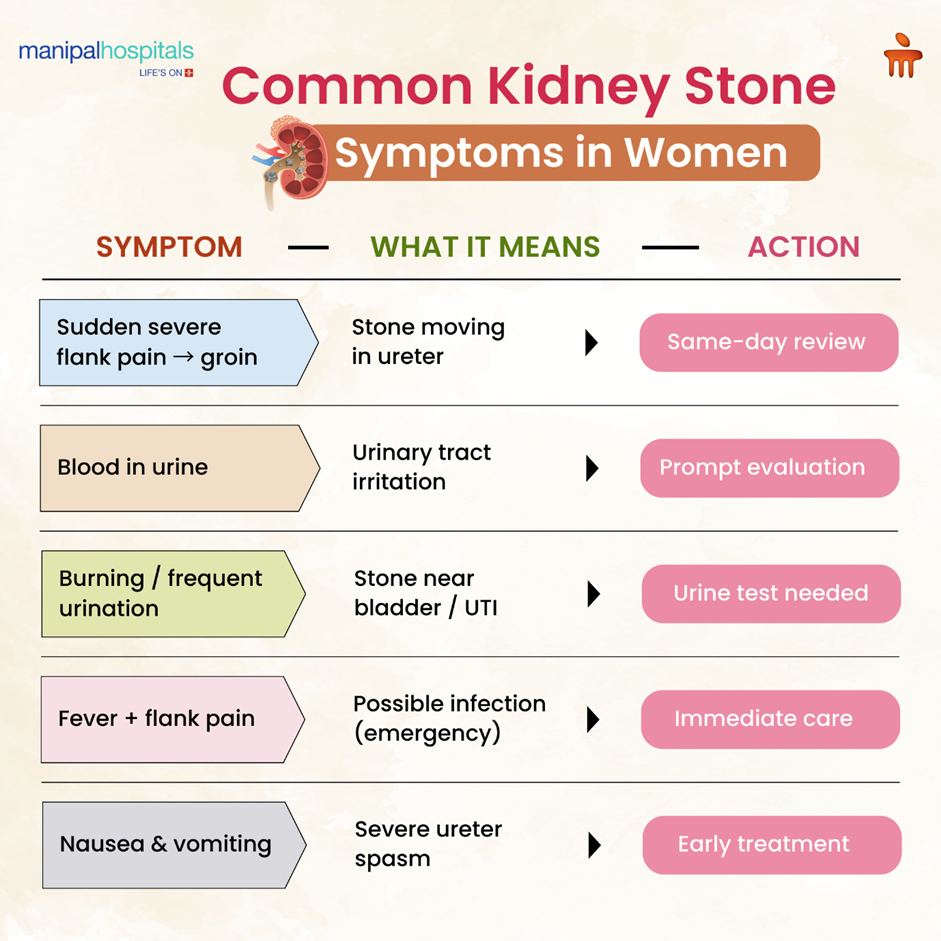
Common Early Signs: What to Look For
Recognising kidney stone symptoms in women early makes a big difference. Common early signs include:
If you experience any of these, especially when several occur together, it is sensible to seek medical assessment quickly.
Why Kidney Stones Happen: Common Causes
Understanding the causes of kidney stones in women can help reduce the risk of recurrence. Several contributing factors increase the likelihood of stone formation.
-
Low fluid intake: Concentrated urine increases the chances of crystal formation.
-
Dietary factors: High salt intake, excessive animal protein, and high-oxalate foods such as spinach and nuts can raise the risk.
-
Metabolic issues: High urinary calcium, low citrate levels, elevated uric acid, or other metabolic abnormalities may promote stone formation.
-
Urinary infections: Certain bacteria can encourage the formation of struvite stones.
-
Hormonal changes and pregnancy: Shifts in hormones and fluid balance can influence stone risk.
-
Genetic predisposition or medical conditions: Conditions such as hyperparathyroidism may increase susceptibility.
How Kidney Stone Pain in Women Typically Feels
Kidney stone pain in women often follows a characteristic pattern, though symptoms can sometimes overlap with other abdominal or gynaecological conditions.
-
Sudden, severe pain: The pain is usually abrupt and intense, often described as coming in waves as the ureter goes into spasm.
-
Shifting pain location: As the stone moves, pain may travel from the flank to the lower abdomen, groin, or even the inner thigh.
-
Atypical presentation: Some women report lower abdominal discomfort or pelvic pain instead of classic flank pain, which is why doctors evaluate both urinary and gynaecological causes.
-
Fever with pain: If fever occurs along with severe pain, it may indicate an infected, obstructed stone and requires emergency medical attention.
Effective pain control and prompt diagnosis are essential, as both severe pain and infection can pose significant health risks.
Diagnosis: What to Expect at the Doctor
Accurate kidney stone diagnosis for women in Mysore typically follows a structured clinical approach to confirm the presence, size, and location of the stone while ruling out other possible causes.
-
History and physical examination: The doctor will assess the description and pattern of pain, urinary symptoms, previous history of stones, and pregnancy status, where relevant.
-
Urine tests: Dipstick testing and microscopic examination help detect blood, infection, and crystals in the urine.
-
Blood tests: These evaluate kidney function (creatinine levels), white blood cell count, electrolytes, and uric acid when indicated.
-
Imaging: A non-contrast CT scan is considered the gold standard for most patients. Ultrasound is preferred during pregnancy or when avoiding radiation exposure.
-
Stone analysis: If a stone is passed naturally or removed during a procedure, laboratory analysis of its composition helps guide long-term prevention strategies.
First-Line Home Care While Arranging Tests
These steps can help manage symptoms temporarily while awaiting medical evaluation:
-
Stay well hydrated: Sip water steadily unless your doctor advises fluid restriction.
-
Use a heat pack: Applying gentle heat to the painful side may provide short-term relief.
-
Take appropriate over-the-counter pain relief: Paracetamol or NSAIDs (if suitable for you) can help. NSAIDs are often effective in reducing ureteral spasm.
-
Limit strenuous activity but try gentle walking: Light movement may sometimes help small stones pass.
-
Save passed stone fragments: Collect any fragments for laboratory analysis to guide prevention.
Do not delay medical attention if the pain becomes severe, fever develops, or urine output significantly decreases or stops.
Treatment Options
Treatment for kidney stones in women depends on the stone’s size, location, severity of symptoms, and whether infection is present.
Conservative Management
-
Observation for small stones: Stones smaller than 5–6 mm often pass naturally with adequate hydration and pain control.
-
Medical expulsive therapy: Alpha-blockers may be prescribed to relax the ureter and help the stone pass more easily.
-
Follow-up imaging: Repeat scans confirm whether the stone has passed or if further intervention is required.
Minimally Invasive Procedures
-
Shock Wave Lithotripsy (SWL): A non-invasive method that uses focused sound waves to break stones into smaller fragments. Suitable for selected stone sizes and locations.
-
Ureteroscopy (URS) with Laser Lithotripsy: An endoscopic procedure used to fragment and remove ureteral and certain kidney stones, offering high success rates.
-
Percutaneous Nephrolithotomy (PCNL): A percutaneous approach used for large or complex kidney stones.
-
Emergency drainage (stent or nephrostomy): Performed when an infected, obstructed system requires immediate decompression before definitive treatment.
Discuss the benefits and risks of each option with a urologist to determine the most appropriate approach for your condition.
Special Considerations for Pregnancy and Women’s Health
Pregnancy alters how the body responds to illness, requiring careful selection of diagnostic and treatment approaches to protect both mother and baby.
-
Ultrasound as the primary diagnostic tool: Ultrasound is preferred during pregnancy as it avoids radiation exposure.
-
Limited medication and imaging options: Many drugs and imaging modalities are restricted during pregnancy, so management prioritises maternal and fetal safety.
-
Multidisciplinary coordination: If intervention is required, urologists work closely with obstetric specialists to ensure safe timing and appropriate technique.
During pregnancy, kidney stones require prompt evaluation and carefully coordinated care to ensure the best outcomes for both mother and child.
Prevention: Reducing the Chance of Recurrence
After a first kidney stone episode, prevention becomes a priority. Identifying the underlying cause and making targeted lifestyle, dietary, and medical adjustments can significantly reduce the risk of recurrence and protect long-term kidney health.
The following measures help lower the chances of future stones:
-
Hydration: Maintain adequate fluid intake to achieve a urine output of more than 2 litres per day.
-
Dietary modifications: Limit salt intake, maintain normal (not low) dietary calcium, reduce excessive animal protein, and adjust high-oxalate foods when appropriate.
-
Medications when indicated: Thiazide diuretics for calcium stones, allopurinol for uric acid stones, and citrate supplements when urinary citrate levels are low.
-
Metabolic evaluation: A 24-hour urine analysis helps identify risk factors and enables personalised treatment planning.
A personalised prevention strategy greatly reduces recurrence risk and supports long-term kidney health.
When to Seek Emergency Care
Certain symptoms require immediate medical attention to prevent serious complications.
Seek emergency evaluation if you experience:
-
Severe, uncontrolled pain that does not improve despite medication.
-
Fever or chills with flank pain, which may indicate an infected and obstructed kidney.
-
Inability to pass urine or signs of reduced kidney function.
-
Pregnancy with severe symptoms requires urgent and carefully coordinated care.
Prompt medical intervention helps prevent sepsis, kidney damage, and other serious complications.
Conclusion
Your kidneys are vital to keeping your body balanced and healthy, and they deserve your attention and care. Detecting the kidney stone symptoms in women early and getting timely medical support is the key to restoring healthy kidney functioning. At Manipal Hospital Mysuru, we offer advanced diagnosis, fluid management, infection control, and dialysis to make the patients feel confident.
For expert evaluation, advanced imaging, and personalised kidney stone care, consult the experienced urology specialist at Manipal Hospitals Mysuru for comprehensive and timely treatment support.
FAQ's
Women affected with kidney stones may experience pain that mimics menstrual pain or UTI. Hence, they may feel the pain is located more toward the lower abdomen or pelvis, which is uncommon when compared to men. In addition, women may also complain of a burning sensation while passing urine and frequent urination.
To diagnose acute kidney injury, doctors measure blood creatinine and urea levels, analyse urine for abnormalities, and use imaging studies to identify possible structural or obstructive problems in the kidneys.
Many small stones pass without intervention, but some become stuck and require procedures. Follow-up imaging confirms passage.
Yes, early detection and prompt treatment can reverse acute kidney injury. Recovery depends on the cause, the person's overall health, and how quickly they get medical care.
Advanced nephrology centres provide complete kidney stone diagnosis for women in Mysore along with comprehensive care for acute kidney injury, supported by modern equipment, dialysis facilities, and experienced kidney specialists.
Share this article on:


.webp "Discomfort While Eating: Causes, Symptoms & Dysphagia Treatment")