What are the different types of angiogram procedures?
-
Coronary Angiogram Procedure: An angiogram of the arteries that bring blood to the heart muscle is called a coronary angiogram. It is performed to diagnose coronary artery disease (CAD) caused by blockages or narrowing of the coronary arteries. Coronary artery disease is most often the primary cause of heart attack. A coronary angiogram helps determine the severity of CAD and is also helpful in determining whether an individual is fit for angioplasty or coronary artery bypass surgery. The procedure also provides vital information on the structure of the heart vessels.
-
Aortic Angiogram Procedure: An aortic angiogram is the angiogram of the aorta, the large artery of the heart that carries blood away from the heart to the rest of the body. An aortic angiogram often reveals a blockage or an aneurysm, where an aneurysm is defined as a weakened or bulged area of the artery. It is also useful in detecting aortic regurgitation (valve leakage between the aorta and the left ventricle)
-
Aortofemoral Angiogram Procedure: Aortofemoral angiogram is also called the angiogram of the leg. This angiogram is performed to visualise the arteries of the abdomen and legs.
-
Carotid Angiogram Procedure: Carotid angiogram is performed to examine the carotid arteries, the major arteries travelling up each side of the neck to supply blood to the brain. Carotid angiograms can show if the arteries are narrowed or blocked. Any blockages in carotid arteries can increase the risk of a brain stroke. The angiogram also detects aneurysms, arteriovenous malformation, and vasculitis (inflamed blood vessels).
-
Cerebral Angiogram Procedure: A cerebral angiogram shows the blood vessels in the brain and helps check for aneurysms, blood clots, blockages, malformations, unusual narrowing, internal bleeding, swelling, or changes due to a tumour.
-
Renal Angiogram Procedure : An angiogram to assess the arteries that carry blood to the kidneys.
-
Peripheral Angiogram Procedure (peripheral arteriography): Peripheral angiogram is performed on the arteries of the arm, legs, and trunk, but does not include the aorta or other arteries of the heart. It is mainly performed to detect peripheral arterial disease (PAD), arteriovenous fistulas, and arteriovenous malformations.
-
Pulmonary Angiogram Procedure: A pulmonary angiogram is performed to evaluate the blood vessels of the lung. It helps in the diagnosis of pulmonary embolism, a condition characterized by blood clots or blockages in the pulmonary arteries (the arteries carrying blood from the heart to the lung).
-
Fluorescein Angiogram Procedure: An angiogram for the vessels of the eyes is called the fluorescein angiogram. It helps evaluate eye conditions such as diabetic retinopathy, macular degeneration, etc. and also helps evaluate the retina before laser therapy.
Advanced Techniques to Improve the Accuracy and Effectiveness of Angiograms:
In cases of complicated angiography cases, where standard angiography may not be completely efficient to reveal the full extent or nature of a blockage, causing problem is stent placing, advanced techniques such as advanced intravascular coronary imaging and calcium modification techniques may be used. These techniques are especially helpful in achieving a more targeted, safer, and successful treatment approach.
Advanced intravascular coronary imaging: These techniques are often used to improve the accuracy and effectiveness of Angiograms. These advanced intravascular coronary imaging include:
-
OCT (Optical Coherence Tomography): OCT uses light waves to take ultra-high-resolution pictures of the inside of the blood vessels. This technique enables doctors to see fine details, such as cholesterol or calcium deposits or how well a stent has been placed.
-
IVUS (Intravascular Ultrasound): This technique uses sound waves to produce real-time images of the blood vessel walls from the inside and help in assessing how narrowed or blocked a vessel is. It guides accurate stent placement during procedures.
-
NIRS IVUS (Near-Infrared Spectroscopy combined with IVUS): This technique helps identify high-risk blockages that may be missed on standard angiograms. The technique combines two technologies to detect both the structure of the artery and the type of plaque (fatty or fibrous).
Calcium Modification Techniques During Coronary Interventions (PCI): In certain cases, the arteries are extremely narrowed or may have hardened calcium deposits, which may cause hindrance in placing a stent using standard techniques. For such cases, calcium modification techniques are used during Percutaneous Coronary Intervention to improve stent outcomes and reduce the risk of restenosis. These techniques include,
-
Rotablation: Rotablation or rotational atherectomy, uses a tiny rotating burr to drill through hard calcium inside the artery. The burr creates a smoother path for the stent, reducing the risk of complications.
-
Orbital Atherectomy: This method breaks down the calcium deposits into tiny particles using an orbiting motion to break it into tiny particles. By sanding down the calcium deposits, the natural shape of the artery is preserved and damage to healthy tissue is minimised.
-
IVL (Intravascular Lithotripsy): IVL, also called shockwave therapy, is a minimally invasive procedure that works on the same principle as kidney stone removal. It uses gentle sonic pressure waves to fracture the hardened calcium. The procedure is very safe and has excellent success rates, especially in elderly patients or those with very hard blockages.
By using these advanced calcium modification techniques, our interventional cardiologists are able to successfully treat complex and heavily calcified coronary artery disease—improving (re-narrowing of the artery).
Checklist: Preparing for Angiogram
-
Follow all the instructions provided by your healthcare provider.
-
You may be asked to not eat or drink anything for fixed hours on the day of the procedure (usually after midnight on the day of the procedure)
-
Inform the health care provider in case you have any kidney problems or allergies to iodine-containing substances, such as contrast dye.
What happens during an Angiogram procedure?
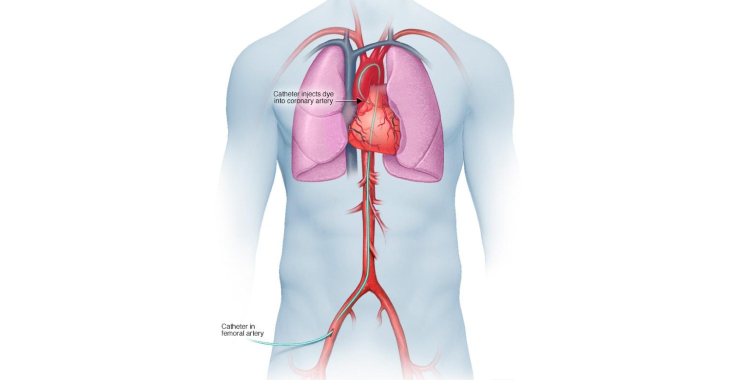
A small amount of discomfort can be expected during the procedure. However, your doctor will inject you with a local anaesthetic that will help you not experience pain during the placement of the catheter. The steps during an angiogram procedure include:
-
The catheter is inserted and guided to the artery being checked.
-
The doctor then injects dye into the artery. You may experience a slightly warm feeling or hot flush spreading over all or part of your body for a few seconds.
-
A series of X-rays are taken as the dye moves through your artery. These X-rays show where the artery is blocked or narrowed and also help determine the degree of deformity, blockage, or narrowing.
-
Once the procedure is over, the catheter is removed.
The whole procedure will take about one hour and can be carried out on an out-patient basis.
What happens after an Angiogram procedure?
After the procedure, you will be kept under observation for at least a few hours until the risk of bleeding is ruled out. You may be allowed to go home in case there are no complications. You will be advised to avoid all strenuous activity for at least the rest of the day to prevent bleeding from the point where the catheter was inserted.
Benefits of Angiogram procedure
-
Precision and Accuracy: The great degree of precision of an angiography procedure is one of its most important advantages. The contrast dye injected makes it possible to precisely identify obstructions, constriction, or abnormalities which is necessary to advise the severity of vascular disorders, this precision is essential.
-
Efficiency and Timeliness: Angiogram procedure is a quick process thereby helping in quick diagnosis. This promptness can save lives in emergency situations, such as when a suspected heart attack or stroke occurs, by guaranteeing that patients receive the right care at the right time.
-
Very Minimal Invasion: Angiogram is less intrusive than open surgical techniques. There is less discomfort, a shorter recovery period, and a lesser chance of problems making this the best option for both diagnosis and intervention.
-
Adaptability in Therapy: Angiogram is used to guide treatment interventions in addition to diagnosing diseases. For example, a stent or balloon angioplasty can be used to restore blood flow after a blockage has been found. It is extremely useful in contemporary cardiology because of its dual function as a diagnostic and therapeutic tool.
Why Choose Manipal Hospitals Bangalore for an Angiogram procedure?
Manipal Hospitals, Bangalore, is a trusted name in healthcare that is well-known for its superior cardiac treatment, top cardiology doctors and diagnostic services. With an outstanding track record of nearly 18,000 angiograms performed annually across Manipal Hospitals Bangalore, we excel in accurately diagnosing even the most complex vascular conditions with precision and care. When you choose Manipal Hospitals for your angiography you are assured of:
-
State-of-the-Art Technology: Latest and most advanced imaging and diagnostic tools that guarantee accurate and precise findings.
-
Experienced Specialists: A team of highly skilled vascular specialists, interventional radiologists, and highly qualified cardiologists committed to providing the greatest treatment.
-
Comprehensive Patient Care: A patient-centred strategy that includes individualised treatment programmes and aftercare.
-
Minimally Invasive Techniques: Cutting-edge methods to guarantee a speedy, painless, and seamless recovery.
-
Emergency Cardiac Care: 24/7 emergency cardiac services with fast-track interventions for patients in critical circumstances.
When it comes to your heart and vascular health, choosing the correct hospital makes all the difference. Manipal Hospitals guarantees that you will receive compassionate, skilled, and top-notch care. Avail the best treatment for Angiogram Procedures in Bangalore now at Manipal Hospitals.
Meet Our Specialists