7 Min Read
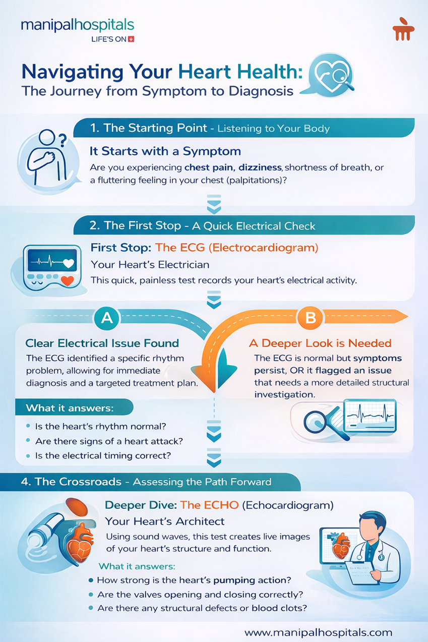
The human heart, a remarkable organ, works tirelessly to sustain life. When concerns about its function arise, understanding the diagnostic tools available becomes paramount. Among the most common and vital cardiac tests are the Electrocardiogram (ECG/EKG) and the Echocardiogram (ECHO). While both are non-invasive and provide invaluable insights into heart health, they measure different aspects and serve distinct purposes. In this blog, let us explore the difference between ECG and echo and help you navigate your diagnostic journey and maintain optimal heart health.
Synopsis
Understanding the Electrocardiogram (ECG/EKG)
An ECG test is a fundamental diagnostic tool that records the electrical activity of your heart. Every beat of your heart is triggered by an electrical impulse. An ECG captures these impulses as they travel through your heart, translating them into waves displayed on a graph or monitor. It’s a quick, painless, and non-invasive procedure, often serving as the first line of investigation for various heart-related symptoms.
How Does an ECG Work?
During an ECG, small adhesive electrodes are attached to specific points on your chest, arms, and legs. These electrodes detect the tiny electrical changes on your skin that arise from the heart muscle's electrophysiological pattern. The machine then amplifies and records this electrical activity, producing a tracing that provides information about your heart rate, rhythm, and the strength and timing of electrical signals as they pass through the heart chambers. This process typically takes only a few minutes.
When is an ECG performed?
An ECG test is commonly performed for a variety of reasons, including:
-
Evaluating symptoms such as chest pain, dizziness, palpitations, or shortness of breath.
-
Detecting an irregular heartbeat (arrhythmia).
-
Diagnosing a heart attack (myocardial infarction) or assessing damage from a previous one.
-
Checking the effectiveness of certain heart medications.
-
Assessing heart health before surgery.
-
Screening for certain heart conditions, especially in individuals with risk factors.
ECGs are instrumental in the rapid diagnosis of heart attacks, with studies showing they can identify acute myocardial infarction with over 90% accuracy when performed promptly. Its simplicity and speed make it an indispensable tool in emergency cardiac care.
Understanding the Echocardiogram (ECHO)
An echo test, or echocardiogram, is a more sophisticated diagnostic tool that uses high-frequency sound waves (ultrasound) to create live images of your heart. Unlike an ECG, which looks at electrical activity, which is a major difference between ECG and echo, an ECHO provides a detailed view of your heart's structure, including its chambers, valves, and major blood vessels, as well as how well it's pumping blood.
How Does an ECHO Work?
During an ECHO, a trained sonographer applies a special gel to your chest and then presses a transducer (a small, hand-held device) against your skin. The transducer emits sound waves that bounce off your heart and create echoes. These echoes are then converted into real-time images and videos that can be viewed on a monitor. This allows cardiologists to observe the heart's movement, measure blood flow, and identify any structural abnormalities.
When is an ECHO performed?
An echo test is typically recommended for:
-
Investigating heart murmurs or unusual heart sounds.
-
Diagnosing and monitoring heart valve problems (e.g., stenosis or regurgitation).
-
Assessing the pumping strength of the heart, particularly in cases of heart failure.
-
Detecting congenital heart defects (heart problems present at birth).
-
Evaluating the size and shape of the heart chambers.
-
Identifying blood clots or tumours within the heart.
-
Assessing the pericardium (the sac surrounding the heart).
Echocardiograms provide a wealth of information, enabling cardiologists to assess heart function with an accuracy exceeding 95% for many structural heart abnormalities. They are crucial for detailed anatomical and functional assessments.
ECG Vs ECHO: Key Differences at a Glance
While both tests are vital for diagnosing heart conditions, understanding the difference between ECG and ECHO is key. They are complementary, offering distinct pieces of the cardiac puzzle.
Below is an electrocardiogram vs echocardiogram comparison table to highlight their unique contributions to heart health:
Difference Between ECG and ECHO
|
Feature |
ECG (Electrocardiogram) |
ECHO (Echocardiogram) |
|
What they Measure |
Electrical activity of the heart (rate, rhythm, electrical timing). |
Physical structure, function, and blood flow (chambers, valves, and muscle strength). |
|
Technology Used |
Electrodes detect electrical impulses. |
Ultrasound waves create moving images. |
|
Information Provided |
Detects arrhythmias, heart attacks, and electrical pathway issues. |
Visualizes heart muscle damage, valve disease, chamber enlargement, and pumping efficiency. |
|
Time Taken |
Typically 5 - 10 minutes. |
Usually 30 - 60 minutes, depending on required detail. |
|
Primary Role |
Quick initial assessment of electrical stability and acute events. |
Detailed structural and functional assessment |
The Role of ECG and ECHO in Cardiac Care at Manipal Hospitals Bangalore
We understand that navigating heart health can be concerning. At Manipal Hospitals Bangalore, our state-of-the-art cardiology department is equipped with advanced diagnostic technologies, including the latest ECG and ECHO tests in Bangalore. Our team of highly skilled cardiologists and allied support teams leverages these tools to provide accurate diagnoses and personalized treatment plans. Our patient-centric approach ensures that you receive comprehensive care, from initial consultation and diagnosis to treatment and ongoing management.
So, if you are experiencing any heart symptoms, book an appointment with our expert cardiologists at Manipal Hospitals today.
FAQ's
No, an ECG is a painless and non-invasive procedure. You might feel a slight tug when the electrodes are removed, but there's no discomfort during the test itself.
An echocardiogram typically takes between 30 and 60 minutes, depending on the type of ECHO performed and the amount of detail required by the cardiologist.
Both ECG and ECHO are very safe procedures with no known risks. They are non-invasive and do not use radiation.
For an ECG, no specific preparation is usually needed, other than avoiding lotions on your chest. For an ECHO, you may be asked to avoid eating or drinking for a few hours before the test, especially if a transoesophageal echocardiogram (TEE) is planned, though a standard transthoracic ECHO often requires no special preparation.
No, an ECG cannot replace an ECHO, and vice versa. They provide different, yet complementary, information about your heart. An ECG assesses electrical activity, while an ECHO visualises the heart's structure and function. Both may be necessary for a comprehensive cardiac evaluation.
You should seek medical advice if you experience symptoms like persistent chest pain, shortness of breath, unexplained fatigue, palpitations, swelling in your legs, or dizziness. Regular check-ups are also crucial, especially if you have risk factors like high blood pressure, diabetes, or a family history of heart disease.
Share this article on: