.webp)
The most common cervical spinal fracture is an odontoid fracture, particularly Type II, which occurs due to trauma or spinal column degeneration. These fractures compromise spinal stability, leading to pain, neurological deficits, and reduced mobility. Surgical intervention is often required for Type II odontoid fractures, as they have a high risk of nonunion despite initial bracing treatment.
Patients prefer anterior odontoid screw fixation because it stabilises the fracture while preserving neck mobility. This minimally invasive technique promotes faster healing and reduces complications associated with traditional fusion methods.
What is Anterior Odontoid Screw Fixation?
Surgical teams use Anterior Odontoid Screw Fixation to stabilize fractures of the odontoid bone by inserting screws through the front of the neck. Surgeons can precisely stabilise the fracture site through this direct approach. The secured fracture allows the body to heal while maintaining normal spine movement. Unlike posterior fusion methods that restrict movement, anterior screw fixation preserves cervical motion, making it ideal for active individuals who need full spine function.
Is Anterior Odontoid Screw Fixation a Major Surgery?
Yes, anterior odontoid screw fixation is a major surgical procedure, but it is minimally invasive compared to other spinal surgeries.
Surgical Procedure Overview:
-
Performed under general anaesthesia – Ensures a pain-free and safe procedure.
-
Small incision at the front of the neck – Provides direct access to the odontoid process with minimal tissue disruption.
-
Insertion of screws through the odontoid – Specialised titanium screws are placed to stabilize the fracture and promote healing.
Despite being a major surgery, its minimally invasive nature allows for quicker recovery and preservation of neck mobility.
What Health Conditions Require Anterior Odontoid Screw Fixation?
Type II Odontoid Fractures
Most patients undergo anterior odontoid screw fixation for Type II odontoid fractures, which break at the base of the odontoid process (dens). These fractures have a high risk of nonunion when treated with external bracing alone. Patients typically choose surgical intervention because the odontoid is essential for spinal stability and head movement. Surgery creates optimal conditions for proper healing and prevents long-term complications.
Minimally Displaced Fractures
This surgical technique works best for odontoid fractures that maintain proper alignment and aren't severely displaced. Anterior screw fixation effectively supports healing while preserving normal spinal function. The procedure is less effective for fractures with extreme displacement or angulation, which may require posterior cervical fusion instead.
Patients with High Functional Demand
People who need neck mobility for work experience major benefits from this procedure. Athletes, manual workers, and other professionals in physically demanding occupations require normal spinal function. Anterior odontoid screw fixation allows patients to maintain cervical movement, enabling them to perform regular activities freely. The procedure is also suitable for elderly patients who want to preserve independence and avoid complications from spinal fusion.
Failure of Conservative Treatment
Surgical intervention becomes necessary when non-surgical treatments fail to heal the fracture. Anterior odontoid screw fixation is needed when X-rays or CT scans show delayed healing, misalignment, or worsening symptoms after treatment with a rigid cervical collar or halo brace. Prompt surgery in these cases prevents additional problems like persistent pain, spinal deformity, and neurological damage.
Indications for Surgery
-
Unstable Type II Odontoid Fractures – Without surgical intervention, these fractures have a high likelihood of nonunion, leading to chronic instability.
-
Neurological Symptoms – If the fracture compresses the spinal cord or nerves, causing weakness, numbness, or coordination issues, surgery is indicated.
-
Severe Neck Pain and Instability – Persistent pain and difficulty maintaining head posture due to fracture instability necessitate surgical stabilisation.
-
Fracture Displacement Greater than 5mm – Displaced fractures have a significantly higher risk of healing failure without surgical fixation, making surgery the preferred treatment option.
Key Aspects of Anterior Odontoid Screw Fixation
Patient Selection & Preoperative Assessment:
Ideal for Anderson–D'Alonzo Type II odontoid fractures with minimal fragmentation, good alignment on traction films and intact transverse ligament. Best suited to physiologically younger patients (typically <65 years) with fractures <6 months old and no significant atlanto‑axial joint displacement.
Anaesthesia & Positioning:
Conducted under general anaesthesia with neuromonitoring. The patient lies flat on a radiolucent table; the head is secured in slight extension (via a Mayfield head clamp or foam supports) to facilitate reduction and optimise fluoroscopic views.
Anterior Cervical Exposure:
A small transverse (skin‑crease) incision is made at the C4–C5 level. The platysma and pretracheal fascia are divided, retracting the carotid sheath laterally and the oesophagus/trachea medially to expose the anterior aspect of C2.
Fracture Reduction Under Fluoroscopy:
Gentle axial traction and neck extension are applied to align the odontoid fragment with the C2 body. Continuous two-plane fluoroscopic imaging (AP and lateral) confirms anatomic reduction before instrumentation.
Guidewire Placement:
A cannulated 1.2–1.6 mm guidewire is advanced through the anterior C2 body, across the fracture line, aiming for the odontoid tip. Precise trajectory planning avoids damage to the spinal canal or vertebral arteries.
Cannulated Lag‑Screw Insertion:
Over the guidewire, a 4.0–4.5 mm partially threaded lag screw is advanced. As the screw is tightened, it creates compression between the fragments, promoting direct bone healing and preserving joint rotation.
Intraoperative Verification:
Final AP and lateral fluoroscopic images ensure proper screw placement, correct length, ideal trajectory and absence of canal or vascular encroachment.
Postoperative Care & Fusion Monitoring:
Patients wear a rigid cervical collar for 6–12 weeks. Early mobilisation is encouraged. Serial radiographs (at 6, 12 and 24 weeks) assess fracture union and screw position. CT may be used if the union is uncertain.
Potential Pitfalls & Complications:
Risk of screw malposition, vertebral artery injury or nonunion. Careful preoperative CT assessment of pedicle and odontoid anatomy, plus meticulous fluoroscopic technique, reduces these risks.
Benefits of Anterior Odontoid Screw Fixation
Preservation of Cervical Motion
The main benefit of anterior odontoid screw fixation is preserving normal neck movement. The procedure stabilises the fracture without restricting cervical flexibility. This is especially valuable for young patients and those who need full neck mobility for daily activities, sports, or work. The technique maintains normal spinal mechanics, reducing strain on adjacent vertebrae and preventing future functional limitations.
Minimally Invasive Approach
Anterior odontoid screw fixation is performed through a small neck incision. The technique requires minimal tissue cutting and muscle disruption, resulting in less surgical pain and faster recovery compared to traditional open spinal surgery. Patients experience less blood loss, lower infection risk, and quicker return to normal activities. The procedure also produces better cosmetic results with minimal scarring.
Faster Bone Healing
Direct screw placement into the odontoid fracture enhances bone healing speed and efficiency. The internal screw maintains proper alignment of the fracture fragments to achieve natural bone fusion. This results in faster recovery and a lower risk of nonunion. The healing time directly affects how long patients need to remain immobilised before regaining independence.
Reduced Risk of Adjacent Segment Degeneration
A major drawback of spinal fusion surgeries is increased pressure on neighbouring vertebrae, leading to accelerated degeneration. Anterior odontoid screw fixation preserves normal movement, reducing stress on adjacent spinal levels. This lowers the risk of developing adjacent segment disease. Patients benefit from better long-term spinal health by avoiding the progression of degenerative changes in the cervical spine.
Will I Need Any Pre-Procedure Investigations?
Before undergoing anterior odontoid screw fixation, patients must undergo comprehensive diagnostic tests to ensure safety and optimal surgical planning:
-
Imaging Tests – X-rays, CT scans, and MRIs provide detailed views of the fracture and spinal alignment.
-
Bone Density Testing – Determines bone strength and suitability for screw fixation, particularly in older patients.
-
Neurological Evaluation – Assesses any spinal cord involvement or nerve compression requiring additional interventions.
-
Cardiac and Pulmonary Clearance – Ensures patients are fit for general anaesthesia and the demands of surgery.
These investigations help the surgical team deliver precise, safe, and effective treatment for odontoid fractures.
What Happens During the Procedure?
1. Patient Preparation and Anaesthesia
Before surgery, patients undergo thorough evaluation, including imaging and pre-operative tests. General anaesthesia is administered to ensure a pain-free experience. The patient is positioned lying flat with the head stabilised to prevent movement during the procedure.
2. Incision and Access
A small horizontal incision is made in the front of the neck, typically at the C4-C5 level. The surgeon carefully separates soft tissues and muscles while avoiding major blood vessels and the esophagus. This anterior approach provides direct access to the fractured odontoid without disturbing the posterior cervical structures.
3. Screw Placement
Under real-time X-ray guidance, a specialised drill creates a pathway through the fractured odontoid. A titanium screw is carefully inserted across the fracture site, ensuring stable fixation. This technique allows the bone to heal naturally while maintaining normal neck mobility.
4. Closure and Recovery
Once the screw is securely placed, the incision is sutured, and a sterile dressing is applied. Patients are monitored in a recovery unit, and post-operative imaging confirms proper screw positioning before discharge.
How long does it take to recover from Anterior Odontoid Screw Fixation?
1. Hospital Stay
Most patients stay in the hospital for 2-3 days, during which pain management and early mobility exercises begin.
2. Return to Light Activities
Within 2-4 weeks, patients can resume light activities, such as walking and household tasks, with some movement precautions.
3. Full Recovery
The healing process typically takes 3-6 months. During this period, the fractured odontoid bone fuses completely, ensuring spinal stability.
4. Physiotherapy and Follow-Ups
A structured rehabilitation program, including physiotherapy, helps restore neck strength and function. Regular follow-up visits with X-rays or CT scans ensure proper bone healing and screw stability.
By following medical advice and post-operative guidelines, patients achieve optimal recovery and regain their normal lifestyle.
Anterior Odontoid Screw Fixation is a highly effective surgical procedure for stabilising odontoid fractures while preserving neck mobility. With minimally invasive techniques, faster recovery, and a lower risk of complications, it is a preferred option for suitable patients.
At MIRSS, spine surgeons use advanced imaging technology for precision and successful outcomes. The comprehensive care approach, including post-surgical rehabilitation, supports optimal recovery.
If you have sustained a cervical spine injury or require specialised spinal care, consult spine specialists for a personalised treatment plan tailored to your needs.
How Will the Doctor(s) Decide if I Am an Eligible Candidate for Anterior Odontoid Screw Fixation?
This procedure is suitable for select patients based on various medical and anatomical factors:
-
Age and Bone Quality – Ideal for younger patients with good bone density, ensuring better screw fixation and healing.
-
Fracture Alignment – Best suited for well-aligned fractures without excessive angulation or displacement.
-
Spinal Stability Assessment – Ensuring ligament integrity is crucial for successful fixation.
-
General Health Condition – Patients must be fit for anaesthesia and surgery, with no severe comorbidities affecting healing.
A thorough assessment by spine specialists determines the suitability of this procedure for each patient.
Are There Any Risks or Side Effects of Anterior Odontoid Screw Fixation?
Like any surgical procedure, Anterior Odontoid Screw Fixation carries some risks, though complications are rare with expert surgical execution.
1. Infection Risk
Although uncommon, infections can occur. Preventive measures, including sterile surgical techniques and post-operative antibiotics, minimise this risk.
2. Hardware Loosening
In rare cases, the titanium screw may loosen or fail to integrate properly with the bone, potentially requiring revision surgery.
3. Dysphagia (Swallowing Difficulty)
Since the procedure involves an anterior neck approach, some patients may experience temporary difficulty swallowing. This usually resolves within a few days to weeks.
4. Nerve Injury
The surgery is performed close to critical nerves, but with precise navigation techniques, the risk of nerve damage is extremely low.
Careful patient selection, experienced surgical hands, and post-operative monitoring significantly reduce the likelihood of these complications.
Why Choose MIRSS for Anterior Odontoid Screw Fixation?
MIRSS is a leading centre for spine surgery, offering advanced treatment for odontoid fractures. The expertise in anterior odontoid screw fixation helps achieve optimal patient outcomes with minimal complications. Here's why MIRSS stands out:
-
Expert Spine Surgery Team – We have a team of highly experienced spine care specialists, robotic spine surgeons, spine anaesthesiologists, and physiotherapists who collaborate to ensure each surgical outcome is successful at the hospital.
-
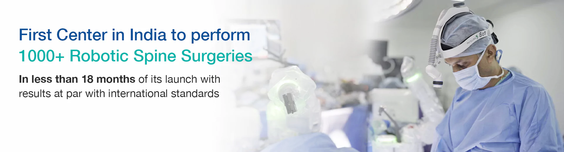
Cutting-Edge Surgical Technology – MIRSS is equipped with 3D imaging and robotic-assisted surgery using Mazor X Stealth Edition Spine Robotics system and O-arm guidance as well as intraoperative navigation systems to visualise the spine in real-time for precise screw placement and enhance surgical accuracy, reducing human error and improving patient outcomes.
-
Comprehensive Pre- and Post-Operative Care – Personalised rehabilitation programs support faster recovery and improved mobility after surgery.
-
Proven Success Rates – High patient satisfaction and superior healing rates highlight the effectiveness of the approach.
With a patient-centered approach and modern technology, MIRSS provides effective care for anterior odontoid screw fixation.