8 Min Read
In the landscape of modern medicine, precise diagnosis is the cornerstone of effective treatment, especially when battling complex diseases like cancer. Among these, the PET CT scan stands out as a revolutionary diagnostic cancer screening tool that combines two powerful imaging techniques into one seamless system, providing unparalleled clarity and detail.
In this blog, let us walk you through this advanced cancer screening technology that has been proven highly effective in improving our chances of the best possible cancer care.
Synopsis
PET CT Scan: A Dual Advantage for Cancer Diagnosis
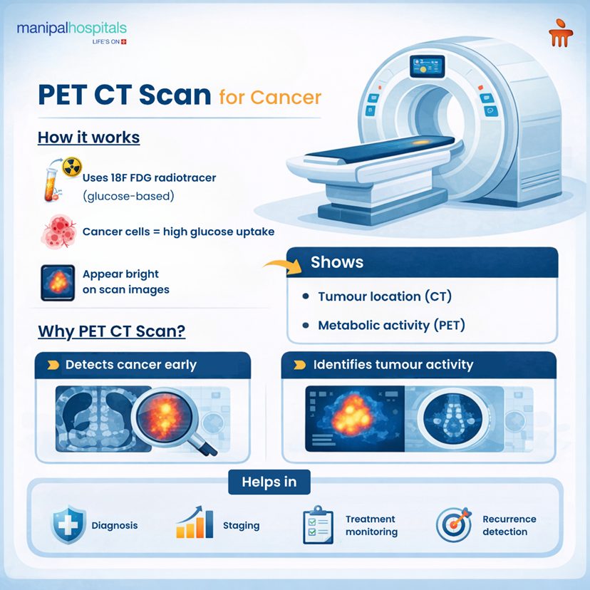
A PET CT scan is a combination of two technologies in one system—Positron Emission Tomography (PET) and Computed Tomography (CT)—where we use a special radiotracer called an 18F FDG molecule. It gives us details of both the structure and functional activity of the body.
Usually, many cancer cells are fast-growing and require glucose molecules for their growth. When the 18F FDG tracer is introduced, these hyperactive cancer cells absorb it in greater amounts. The PET CT machine captures the radiation emitted by these cells and translates it into detailed images. This allows our specialists to visualize not just the location and size of a tumour (structure) but also its metabolic activity (function), often revealing abnormalities much earlier than conventional imaging methods.
This sophisticated imaging technique plays a critical role in the diagnosis and monitoring of various cancers, including lymphomas, lung cancer, breast cancer, and gastrointestinal tract cancers. The ability to peer into the functional aspects of cells makes the PET CT scan for cancer an indispensable tool in modern oncology.
Who Can Benefit from a PET CT Scan?
Most individuals across various age groups can safely undergo a PET CT scan.
-
It is generally safe with minimal side effects.
-
Special instructions are provided for lactating women.
-
Pregnant women are typically advised against it.
-
Extremely overweight patients might have some practical difficulties with the scanner bed, which will be discussed during consultation.
The PET CT Journey: Guidance to Follow
Preparing for and undergoing a PET CT scan is a carefully managed process designed to ensure patient comfort and accurate results. Here’s a detailed overview of what you can expect:
Before Your Arrival: Preparing for the Scan
-
Fasting: An overnight fast or at least 6 hours of fasting is required before the scan to ensure accurate tracer uptake.
-
Activity Restrictions: Avoid strenuous exercise or deep tissue massage for 48 hours prior to the scan, as muscle activity can alter FDG uptake.
-
Medication & Water: You can generally take plain water and your regular medications, unless advised otherwise by your doctor.
-
Inform Doctors About Concerns: It's crucial to inform your doctor about any possibility of pregnancy, if you are breastfeeding, or if you have any internal medical devices.
-
Valuables: For your convenience and safety, please leave your jewellery and other valuables at home.
During Your Visit: The Scanning Process
-
Explanation: Upon arrival, a technologist will explain the procedure in detail, addressing any concerns you may have.
-
Tracer Injection: You will receive a small amount of the intravenous 18F FDG radiotracer injection, usually into a vein in your arm.
-
Rest & Absorption: Following the injection, you will rest quietly for about 45 to 60 minutes in a calm and relaxed environment. This allows the tracer to distribute throughout your body and be absorbed by the target cells.
-
Image Acquisition: Using our advanced PET CT machine, images of your body will be acquired. You will lie on a comfortable table that slides into the scanner.
After Your Examination: Post-Scan Care
-
Infant Contact: To minimize radiation exposure, avoid close contact with infants for 10 hours after the scan.
-
Breastfeeding Mothers: Do not breastfeed for 24 hours post-injection. It is advisable to pump and discard breast milk during this period.
-
Proximity to Children/Pregnant Women: Maintain a distance of 6-10 feet from young children and pregnant women for 6-10 hours after the injection.
-
Hydration: Drink plenty of fluids to help flush the radiotracer from your system.
The PET CT Procedure: A Step-by-Step Overview
Injection
-
A small amount of the 18F FDG radioactive molecule is injected into a vein, typically in your arm, through an intravenous cannula. This tracer is essential for highlighting areas of high metabolic activity.
Absorption
-
Patients then rest quietly for about one hour. During this time, the 18F FDG molecule distributes throughout the body, with a higher concentration absorbed by cells with increased energy demands, such as cancer cells or inflamed tissues.
Technology
-
The 18F FDG molecule is nothing but tagging a radioactive substance with a glucose-like molecule. Since cancer cells consume glucose at a much higher rate than normal cells, this glucose-like molecule with its radioactive tag is preferentially taken up by tumour cells. This distinct uptake makes the tumours appear bright on the PET scan images once they are acquired.
Scanning
-
The patient lies on a table without moving, which is crucial for clear imaging. The table then moves slowly into the scanner, which detects the radiation emitted from the body, particularly from areas where the FDG tracer has accumulated.
Data Processing
-
Post-scan, advanced reconstruction algorithms process the collected data. The scanner combines the functional data (metabolic activity) from the PET scan with the anatomical data (structural information) from the CT scan to generate a detailed, comprehensive 3D map using advanced reconstruction algorithms.
What Can a PET CT Scan Detect?
Cancer: One of the primary uses of a PET CT scan is in oncology. Increased 18F FDG tracer uptake is often a strong indicator of malignant tumours. This powerful diagnostic tool assists in several critical aspects of cancer management:
-
Diagnosis
-
Disease staging
-
Monitoring of treatment response
-
Detects early recurrence
-
To identify a suitable lesion for biopsy
-
For radiation therapy planning
Inflammation and Infection: Beyond cancer, a PET CT scan is also invaluable in identifying and characterizing inflammatory or infected tissues. These tissues, like cancer cells, often exhibit increased metabolic activity and sugar consumption, leading to higher FDG uptake. It can detect conditions such as:
-
Tuberculosis
-
Sarcoidosis
-
Certain viral infections
-
Chemotherapy-related drug-induced lung infections
-
Fever of unknown origin
Key Benefits of PET CT Scan
A PET CT scan shows functional activity (metabolism) rather than just structure. It helps us to detect the disease earlier than conventional images.
-
It gives both metabolic and anatomical detail in one scan, reducing the need for multiple separate scans.
-
The test can quickly and accurately assess whether treatments like chemotherapy and targeted therapies are working, allowing doctors to modify treatment strategies as needed, optimizing patient outcomes.
-
PET CT can sometimes help avoid unnecessary biopsies or other invasive diagnostic procedures, improving patient comfort and reducing risks.
Conclusion
The synergy of Positron Emission Tomography (PET) and Computed Tomography (CT) in a single scanner provides an incredibly comprehensive view. While CT offers detailed anatomical images, showing the size, shape, and location of organs and tissues, PET reveals the metabolic activity within these structures. This combination is particularly crucial in oncology, as it allows doctors to distinguish between benign and malignant lesions, accurately stage cancer, and assess the effectiveness of ongoing treatments.
At Manipal Hospital Kanakapura Road, we are committed to leveraging such cutting-edge technology to enhance cancer evaluation, ensuring our patients receive the most accurate diagnosis and personalized treatment plans. Our facilities, including those offering PET-CT scans in Kanakapura Road, Bangalore, are equipped with state-of-the-art machines and staffed by highly skilled nuclear medicine specialist and radiologists.
FAQ's
Yes, a PET CT scan is a generally safe cancer screening test. The amount of radiation exposure is carefully controlled and considered safe for diagnostic purposes. The radioactive tracer has a very short half-life and quickly leaves the body.
The entire process, from arrival to completion, typically takes about 2-3 hours. This includes preparation, tracer injection, absorption time (45-60 minutes), and the actual scan, which lasts around 20-30 minutes.
The procedure is generally painless. You might feel a slight prick during the intravenous injection of the tracer. You will be asked to lie still, which some m
You must fast for at least 6 hours (or overnight) before your scan. Only plain water and essential medications are usually allowed. Your doctor or the imaging center will provide specific instructions.
A CT scan provides detailed anatomical images (structure), while a PET scan shows metabolic activity (function) at the cellular level. A PET CT scan combines both to provide a comprehensive picture, enhancing diagnostic accuracy, especially in cancer evaluation.
Share this article on: